
Petfood: how to prevent insect infestation
Insect contamination of petfoods is a potential problem in tropical countries; this paper offers an overview of the situation and how the risks can be minimized
This article contains images that may offend the sensibilities of children.
Issue number 33.2 Other Scientific
Published 20/10/2023
Also available in Français , Deutsch , Italiano , Português , Español and ภาษาไทย
OA in cats remains underdiagnosed and undertreated, despite its widespread prevalence; this paper looks at how we can surmount the challenge of early diagnosis, leading to better treatment interventions.

Osteoarthritis (OA) is a highly prevalent condition within the feline population, but often goes undetected.
Radiography and current magnetic resonance imaging protocols have limited use in the diagnosis of early or mild feline OA.
Computed tomography is to date reported to be the most sensitive tool for diagnosis of early degenerative changes associated with feline OA.
Other diagnostics such as arthroscopy show promise for the future diagnosis and treatment of feline OA, and warrant further investigation.
Osteoarthritis (OA) is estimated to occur in 61-99% of cats based on radiographic studies 1,2,3 making it one of the leading causes of chronic pain in this species. The condition is characterized by progressive degradation and loss of articular cartilage (Figure 1), related to altered intrinsic mechanisms of the cartilage and influenced by changes in other intra-articular tissues such as the synovium, the subchondral bone and the menisci. The most commonly affected appendicular joints in cats include the elbow, hip, stifle, and hock joints 1,3,4.
Despite the high prevalence, OA in cats remains underdiagnosed and undertreated. Several reasons for this have been postulated, including:
The challenges in diagnosing feline OA only become more marked when considering our desire to make an early diagnosis or detect minor changes, which are critical in facilitating early intervention. Prompt identification will have the greatest potential for providing effective management of OA, since it provides an opportunity to initiate an appropriate, tailored long-term treatment plan and disrupt the progressive vicious cycle of synovial joint deterioration 5. In this article we will address recent advances in the use of clinical metrology instruments, goniometry, diagnostic imaging and arthroscopy which will facilitate achieving that critical early diagnosis.

Figure 1. Intraoperative photograph of a feline stifle with osteoarthritis secondary to medial patellar luxation. Note the cartilage degeneration and loss, both within the trochlear groove and over the medial trochlear ridge on the left of the image.
© Karen Perry
Due to the aforementioned challenges, owner-reported behavioral signs remain the best assessment tool of feline chronic pain in the clinical setting, and therefore represent a critical component of the feline OA work-up. Information about specific behaviors is collected using clinical metrology instruments (CMIs), quality of life (QoL) or health-related QoL (HRQoL) questionnaires. These instruments are constructed based on rigorous research to identify and validate key behaviors that are indicative of pain or QoL, and generally include questions pertaining to mobility, ability and willingness to perform activities, sociability and self-care (eating or grooming). The main goals of such CMIs are to determine when OA-associated pain is present and to detect treatment-associated improvement.
Several clinical metrology instruments, quality of life and health-related quality of life questionnaires have been published for use in cats with chronic painful conditions. These include, but are not limited to:
Additionally, CMIs that are more specific to the individual cat have also been developed, e.g., the Client-Specific Outcome Measures CMI. These questionnaires are based upon distinct activities that the owner feels are problematic for their particular cat, however to date there is no evidence to indicate whether these individualized scales present any benefit over a standardized CMI.
Currently, the FMPI is the most developed CMI available, but it does take some time to complete it. In an effort to provide a quick, easy and practical tool for screening of cats with OA-associated pain and increase owner-awareness of behaviors that may be affected by the condition, a truncated version of the FMPI was recently developed 8. Interestingly, this abridged version did not change the accuracy in screening when compared to its parent version; there were no significant changes in sensitivity, specificity, positive predictive value, or negative predictive value. This Feline Musculoskeletal Pain Screening Checklist (Feline MiPSC) can be used as a starting point for discussion of feline OA with owners and to check the need for further veterinary investigation. It is comprised of six items asking if a specific activity can be performed normally or not, all with Yes/No answers:
If any question is scored as a “no” (i.e., the activity is not normal), this should prompt further evaluation 8. As such, the Feline MiPSC can be used as a screening tool, whereas other tools such as the FMPI or Montreal Instrument for Cat Arthritis Testing might be used for monitoring treatment efficacy 8.
A question remains regarding when the Feline MiPSC should start to be used as a screening tool. While an age-associated increase in OA has been demonstrated in cats 12, the condition is certainly not isolated to older patients. In one study, 34% of cats with a mean age of 6.5 years were reported to have radiographic signs of OA 13, and infrequently cats as young as one year of age show radiographic evidence of change. Given that this screening tool has the potential to not only increase veterinarians’ ability to screen for OA, but also to increase awareness of OA among cat owners, it is the opinion of the authors that the Feline MiPSC should form a routine component of every feline wellness examination, regardless of patient age. Studies have shown wide gaps between the responses of owners who are informed about OA and those who are not when it comes to assessing behavior changes associated with OA 8. Given that cats are most likely to perform their normal behaviors at home, rather than in the clinic environment, improving owner education and engagement through the use of the Feline MiPSC may render owners more likely to detect altered behaviors at home, which will undoubtedly allow diagnoses to be made earlier.
Lauren M. Meneghetti
While gait assessment and orthopedic examinations may be challenging in some cats, with a good understanding of feline behavior and excellent handling skills it is possible to leverage the cat’s unique characteristics to increase compliance. A full description of a feline orthopedic examination is outside the scope of this article, however, some key points which may facilitate an early diagnosis will be highlighted.
The presence of crepitus, joint effusion, joint thickening and a pain response during palpation increases the likelihood that a joint will have radiographic evidence of OA 14. However, the sensitivity and positive predictive value associated with these tests are low, meaning that radiographic OA cannot be diagnosed with certainty based on palpation alone. Interestingly, the specificity and negative predictive value of these tests are higher, indicating that an absence of orthopedic examination findings of pain, crepitus, effusion and thickening can be used to rule out OA with a high degree of certainty 14. Age has been shown to affect the sensitivity and specificity of these tests; as cats increase in age, there is a closer association between the presence of these findings and the presence of radiographic OA. Unfortunately, this renders these tests less useful for the detection of early OA.
One aspect of orthopedic examination that is frequently overlooked is the use of goniometry. Degenerative changes within joints inhibit normal range of motion (ROM), therefore ROM measurements through goniometry can be utilized to help diagnose OA and monitor its progression. Goniometry measurements (Figure 2) are easy to perform and show no clinically significant difference between measurements taken in sedated or non-sedated cats 14,15. Table 1 describes the placement of the goniometer and the normal ranges of motion for each respective joint (averaged between non-sedated and sedated measurements) 15. Cats with increased ROM have been associated with decreased odds of radiographic OA being present, indicating that normal ROM may have value in ruling out the presence of OA; conversely, a decreased ROM has been associated with the presence of radiographic OA 14. The use of goniometry is of particular interest when considering early diagnosis of OA, because the relationship between ROM and presence of radiographic OA does not appear to be affected by age 14.

Figure 2. Goniometry of the left tarsus being performed in a non-sedated cat.
© Karen Perry
Table 1. Details regarding placement of the goniometer for measurement of joint range of motion of the carpus, elbow, shoulder, tarsus, stifle and coxofemoral joint. The normal ranges of motion for each respective joint (averaged between non-sedated and sedated measurements) are also detailed 15.
| Joint and placement of goniometer | Angle in flexion (Mean (95% CI)) |
Angle in extension
(Mean (95% CI))
|
|---|---|---|
| Carpus: Long axis of metacarpal bones III/IV and longitudinal axis of the antebrachium | 22˚ (21-23) | 198˚ (196-199) |
| Elbow: Longitudinal axis of the antebrachium and longitudinal axis of the humerus | 22˚ (22-23) | 164˚ (162-165) |
| Shoulder: Longitudinal axis of the humerus and spine of the scapula | 32˚ (31-32) | 165˚ (162-168) |
| Tarsus: Long axis of metatarsal bones III/IV and longitudinal axis of the tibia | 21.5˚ (21-22) | 167.5˚ (166-170) |
| Stifle: Longitudinal axis of the tibia and longitudinal axis of the femur | 24˚ (24-25) | 164˚ (163-165) |
|
Coxofemoral joint: Longitudinal axis of the femur and a line parallel to a line joining the tuber sacrale and tuber ischiadicum over the greater trochanter
|
33˚ (32-33) | 165˚ (163-167) |
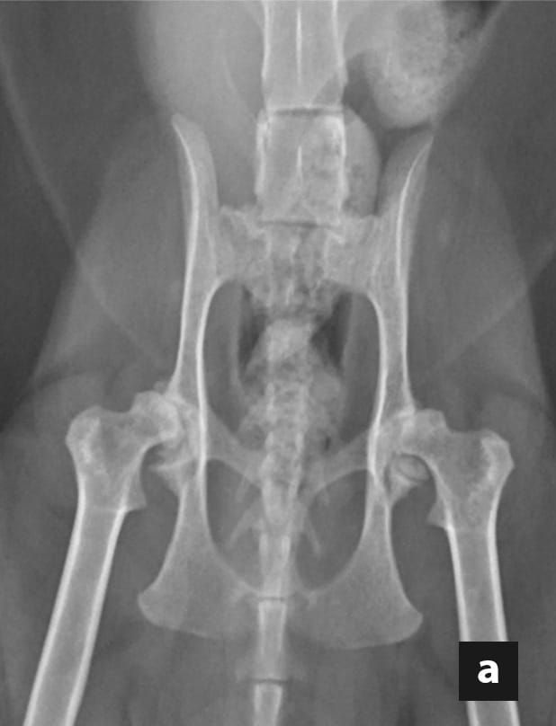
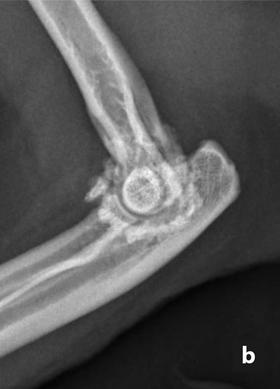
Due to the wide availability and perceived cost effectiveness, radiography remains the most commonly used method for OA diagnosis in cats, despite widespread acceptance that it will only detect severe or advanced OA 16. Radiographic changes appreciated when diagnosing feline OA include the presence of osteophytes, increased subchondral bone opacity, soft tissue and intra-articular mineralizations, soft tissue thickening, and synovial effusion (Figure 3). When degenerative changes are appreciated on radiographs, histologic degenerative changes confirm the presence of OA with a very high specificity 16.
Radiography is a two-dimensional imaging modality, meaning that any changes that are not on, or close to, the edge of skeletal structures are summated on other skeletal structures and may not be visible 16. Additionally, imaging of cartilage using radiography is impossible and radiographic findings do not correlate well with cartilage degeneration; one study demonstrated up to 71% of feline stifle joints with macroscopic evidence of OA did not have associated lesions on radiographs 17. These weaknesses limit the usefulness of radiography for detecting early or mild OA. Indeed, a recent study reported that radiography was not able to detect any joint that was diagnosed with mild OA histologically 16.
Despite these limitations, radiography is often the only imaging modality available and therefore interpretation must be optimized; an awareness of where to look and what to look for will facilitate making an early diagnosis. The development of osteophytosis may be slower in cats than in dogs 2,4 and other radiographic features, including other forms of new bone formation, may be of greater significance 17. While osteophytes are often considered the key radiographic feature of OA, this only appears to be the case for the feline coxofemoral joint 17,18. For the elbow, tarsus and stifle joints, the most common radiographic features are actually joint-associated mineralizations, tarsometatarsal dorsal bone proliferation and intra-articular mineralizations respectively 17. Radiographs of feline joints should be carefully evaluated for these other forms of new periarticular bone in addition to careful assessment for osteophyte formation. Additionally, the limitations of radiography in early diagnosis of OA should always be considered; where OA is suspected but radiographic signs are absent, more sensitive diagnostic modalities should be pursued.
 |
 |
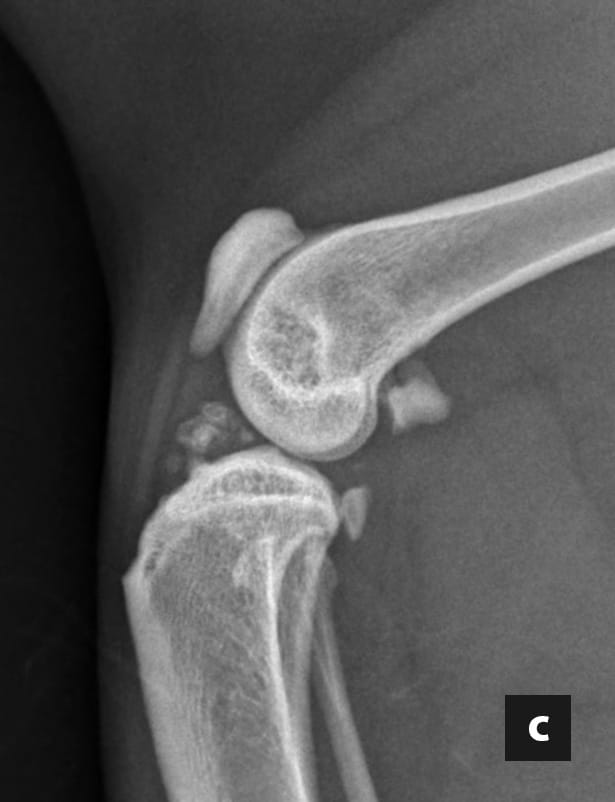 |
Figure 3. Representative images demonstrating the radiographic changes commonly associated with feline osteoarthritis. On the ventrodorsal view of the hips (a), osteophyte formation can be appreciated at the craniodorsal acetabular margins in addition to remodeling of the femoral heads. In the mediolateral view of the elbow (b) increased subchondral bone opacity can be appreciated in the peritrochlear region in addition to periarticular osteophyte formation. On the mediolateral view of the stifle (c) intra-articular mineralization and synovial effusion can be appreciated.
© Karen Perry
Due to multiplanar three-dimensional characteristics, computed tomography (CT) has significant advantages over radiography for the assessment of joints for OA. This is particularly relevant when considering our desire to make an early diagnosis and appreciate mild osteoarthritic changes. Two recent studies have confirmed that this has relevance in the diagnosis of feline elbow and coxofemoral OA specifically 16,18; indeed, CT was able to detect very early stages of disease, even before macroscopic cartilage changes could be detected 18. Histopathologically confirmed OA was detected in all the areas noted to have lesions on CT, indicating high specificity for diagnostic accuracy 18. Additionally, the characteristics of the osteophytes observed on CT may be of assistance in determining condition chronicity; rounded osteophytes appear to be associated with more severe or chronic disease, while spur-shaped and sclerotic, pointed osteophytes appear to be a sign of early OA 18.
As noted above with radiography, looking for specific findings is likely to result in earlier diagnoses being made than when the entire scan is subjectively evaluated. A recent study evaluating the feline elbow demonstrated that while a CT scan did improve sensitivity and specificity for detecting OA in comparison to radiography, subjective grading alone still resulted in a low sensitivity for detecting mild histologic OA 16. However, specific measurement of spurs on the lateral margin of the anconeal process on CT images (Figure 4), and use of a threshold of greater than or equal to 0.5 mm for these measurements, improved this to a moderate sensitivity and high specificity 16.
Recent literature and the authors’ clinical experience indicates that CT can be used in order to make a diagnosis of OA in the earlier and less severe stages of the condition, and this option should be considered in cases where radiographs do not result in a definitive diagnosis. Careful and systematic evaluation of the CT, including the use of specific measurements, are recommended to increase the sensitivity of this imaging modality. While radiography is often considered more cost-effective than a CT scan, it is quick and simple to perform a full body CT scan in cats due to their small size, allowing every joint to be evaluated. This may be considered advantageous in patients where multiple joints are affected based upon orthopedic examination, or in cases where patient disposition impedes a full orthopedic examination and therefore localization of pain.

Figure 4. Frontal plane CT image of the right elbow of an 11-year-old cat presenting with forelimb lameness associated with elbow osteoarthritis. Note the moderate spur formation on the lateral margin of the anconeal process. Measurement of these spurs increases both sensitivity and specificity of computed tomography for detection of mild osteoarthritis in the feline elbow.
© Karen Perry
Magnetic resonance imaging (MRI) has been studied for use in the diagnosis of orthopedic disease due to its superior ability to evaluate soft tissue structures such as cartilage. It is well established that cartilage damage precedes bony changes such as osteophytosis and sclerosis that are routinely evaluated using radiography and CT in patients suffering from OA 19. In theory, the ability to identify cartilage lesions prior to bony changes would make MRI an ideal diagnostic tool for OA in terms of facilitating earlier diagnoses; indeed, in dogs, horses and people, MRI is more sensitive than radiography for assessing OA structural changes including cartilage lesions, osteophytosis, joint effusion and synovial thickening.
MRI has also been used to investigate hip dysplasia and OA in cats in one pilot study 20. While osteophytes and sclerosis were found using both radiography and a 1.5 Tesla MRI, one cat that had no signs detectable with radiography had several OA lesions identified on MRI, including bilateral osteophytosis, joint effusion and thinning of the articular cartilage 20. Additionally, in two cats with OA, MRI revealed bone marrow lesions in the femoral head that were not detected using radiography 20. An additional study carried out high field (4.7 Tesla) MRI of normal feline hip joints and concluded that while the femoral and acetabular subchondral bone could be well visualized, the articular cartilage was more challenging to assess, which would potentially limit the usefulness of this imaging modality to detect the earlier stages of OA 21. Further investigations are certainly warranted, as it is possible that altered patient positioning or alternative sequences may improve the diagnostic potential of this technique, but currently, the additional cost and requirement for general anesthesia associated with this imaging modality seem difficult to justify.
Karen L. Perry
Arthroscopy is widely used in humans, dogs, and horses for the diagnosis and treatment of multiple joint ailments, but its adoption in cats has been slower, likely due to the increased technical difficulty in performing arthroscopy in small feline joints, concern regarding the potential for iatrogenic damage, and the price and availability of the necessary equipment. In recent years, as experience with arthroscopy has increased and smaller instrumentation has become widely available, the use of the modality in cats has also expanded, with many anecdotal reports of success, but there remains a paucity of data regarding its use within peer-reviewed veterinary literature.
Arthroscopy is considered to be the gold standard diagnostic tool for evaluating joint cartilage as evidenced by its superior sensitivity in comparison to radiographs, CT and MRI. Additionally, arthroscopy can be therapeutic in addition to diagnostic; good results have been reported in cats following arthroscopic debridement of osteochondritis dissecans lesions affecting the femoral condyle 22 and after removal of mineralizations within the elbow joint 23. While inherently more invasive than the aforementioned imaging techniques, the development of small diameter needle arthroscopes which can be used on an outpatient basis under sedation may result in increased use of this diagnostic modality. The superior visualization of articular cartilage that is possible with arthroscopy has the potential to facilitate earlier diagnosis of OA in cats in the future.
Other areas of interest in the attempt to facilitate an early diagnosis of feline OA include quantitative sensory testing, accelerometry, force-plate analysis, measurement of serum cytokines, and differential gene expression 24,25,26,27. At this time, these tests have limited value within the clinical setting for diagnosis of OA; they show differences between normal cats and those with known OA-associated pain, but cut-off values for these tests are lacking and commercial availability is scarce. However, further research in these areas is considered likely to render them useful in both diagnosis and treatment efficacy monitoring in the future.
It is imperative that clinicians realize two fundamentals; firstly, that the sooner osteoarthritis is diagnosed in cats the sooner appropriate treatment can be administered; and secondly, that radiography is not the only option for diagnosis of feline osteoarthritis, and indeed that other options offer superior diagnostic abilities. With the increasing availability of imaging modalities such as CT, MRI and arthroscopy, there are opportunities to improve the detection of early OA lesions. Moreover, the use of questionnaires, coupled with goniometry, should be encouraged to facilitate matters.
Lascelles BDX, Henry 3rd JB, Brown J, et al. Cross-sectional study of the prevalence of radiographic degenerative joint disease in domesticated cats. Vet. Surg. 2010;39:535-544.
Hardie EM, Roe SC, Martin FR. Radiographic evidence of degenerative joint disease in geriatric cats: 100 cases (1994-1997). J. Am. Vet. Med. Assoc. 2002;220:628-632.
Kimura T, Kimura S, Okada J, et al. Retrospective radiographic study of degenerative joint disease in cats: Prevalence based on orthogonal radiographs. Front. Vet. Sci. 2020;7:article 138.
Clarke SP, Bennett D. Feline osteoarthritis: a prospective study of 28 cases. J. Small Anim. Pract. 2006;47:439-445.
Cachon T, Frykman O, Innes JF, et al. Face validity of a proposed tool for staging canine osteoarthritis: Canine OsteoArthritis Staging Tool (COAST). Vet. J. 2018;125:1-8.
Freeman LM, Rodenberg C, Narayanan A, et al. Development and initial validation of the Cat Health and Wellbeing (CHEW) questionnaire: a generic health related quality of life instrument for cats. J. Feline Med. Surg. 2016;18(9):689-701.
Benito J, Hansen B, Depuy V, et al. Feline musculoskeletal pain index: responsiveness and testing of criterion validity. J. Vet. Intern. Med. 2013;27:474-482.
Enomoto M, Lascelles BDX, Gruen ME. Development of a checklist for the detection of degenerative joint disease-associated pain in cats. J. Feline Med. Surg. 2020;12:1137-1147.
Tatlock S, Gober M, Williamson N, et al. Development and preliminary psychometric evaluation of an owner-completed measure of feline quality of life. Vet. J. 2017;228:22-32.
Klinck MP, Gruen ME, del Castillo JRE, et al. Development and preliminary validity and reliability of the Montreal instrument for cat arthritis testing, for use by caretaker/owner, MI-CAT(C), via a randomised clinical trial. Appl. Anim. Behav. Sci. 2018;200:96-105.
Bennett D, Morton C. A study of owner observed behavioural and lifestyle changes in cats with musculoskeletal disease before and after analgesic therapy. J. Feline Med. Surg. 2009;11:997-1004.
Freire M, Meuten D, Lascelles D. Pathology of articular cartilage and synovial membrane from elbow joints with and without degenerative joint disease in domestic cats. Vet. Pathol. 2014;51:968-978.
Clarke SP, Mellor D, Clements DN, et al. Prevalence of radiographic signs of degenerative joint disease in a hospital population of cats. Vet. Rec. 2005;157:793-799.
Lascelles BDX, Dong Y-H, Marcellin-Little DJ, et al. Relationship of orthopedic examination, goniometric measurements, and radiographic signs of degenerative joint disease in cats. BMC Vet. Res. 2012;8:10.
Jaeger GH, Marcellin-Little DJ, DePuy V, et al. Validity of goniometric joint measurements in cats. Am. J. Vet. Res. 2007;68:822-826.
Ley CJ, Leijon A, Uhlhorn M, et al. Computed tomography is superior to radiography for detection of feline elbow osteoarthritis. Res. Vet. Sci. 2021;140:6-17.
Freire M, Robertson I, Bondell HD, et al. Radiographic evaluation of feline appendicular degenerative joint disease vs. macroscopic appearance of articular cartilage. Vet. Radiol. Ultrasound 2011;52:239-247.
Ley C, Ramer G, Leijon A, et al. Acetabular margin changes in feline hip joints – Implications for radiographic diagnosis and development of osteoarthritis. Res. Vet. Sci. 2021;137:243-251.
Libicher M, Ivancic M, Hoffmann M, et al. Early changes in experimental osteoarthritis using the Pond-Nuki dog model: technical procedure and initial results of in vivo MR imaging. Eur. Radiol. 2005;15:1504-1504.
Guillot M, Moreau M, d’Anjou MA, et al. Evaluation of osteoarthritis in cats: novel information from a pilot study. Vet. Surg. 2012;41:328-335.
Kamishina H, Miyabayashi T, Clemmons RM, et al. High field (4.7 T) magnetic resonance imaging of feline hip joints. J. Vet. Med. Sci. 2006;68:285-288.
Bright SR. Arthroscopic-assisted management of osteochondritis dissecans in the stifle of a cat. J. Small Anim. Pract. 2010;51:219-223.
Staiger BA, Beale B. Use of arthroscopy for debridement of the elbow joint in cats. J. Am. Vet. Med. Assoc. 2005;226(3):401-403.
Ashwell M, Freire M, O’Nan AT, et al. Characterization of gene expression in naturally occurring degenerative joint disease-associated pain. Vet. J. 2019;243:42-47.
Guillot M, Taylor PM, Rialland P, et al. Evoked temporal summation in cats to highlight central sensitization related to osteoarthritis-associated chronic pain: A preliminary study. PLoS One 2014; 9(5): e97347. https://doi.org/10.1371/journal.pone.0097347
Gruen ME, Alfaro-Cordoba M, Thomson AE, et al. The use of functional data analysis to evaluate activity in a spontaneous model of degenerative joint disease associated pain in cats. PLoS One 2017;12(1): e0169576. https://doi.org/10.1371/journal.pone.0169576
Gruen ME, Messenger KM, Thomson AE, et al. Evaluation of serum cytokines in cats with and without degenerative joint disease and associated pain. Vet. Immunol. Immunopathol. 2017;183:49-59.
Lauren M. Meneghetti
Dr. Meneghetti gained her Doctorate of Veterinary Medicine degree at the Cummings School of Veterinary Medicine at Tufts University in 2020 Read more
Karen L. Perry
Dr. Perry completed her residency in small animal surgery at The Royal (Dick) School of Veterinary Studies, Edinburgh in 2010 Read more
Insect contamination of petfoods is a potential problem in tropical countries; this paper offers an overview of the situation and how the risks can be minimized
Mistakes happen to all of us in veterinary practice; this article looks at how different people react in different ways when things go wrong and – importantly – discusses how we can best cope with mistakes.
Cats are living longer and better lives; how can we ensure that the healthcare we offer them is optimal? This article offers some hints.
House soiling in cats is an all-too-common problem for many owners; this paper offers a holistic approach to help clinicians advise their clients