
Perianal pruritus in the dog
Anal and perianal pruritus can be a real nuisance and requires careful diagnosis as there are several etiologies.
Issue number 25.2 Other Scientific
Published 03/05/2023
Also available in Français , Deutsch , Italiano and Español
Otitis externa can be frustrating for both the owner and the clinician, as treatment requires a lot of effort, often for a protracted period of time. This article details the minimum information that should be provided to the cat and dog owner when the problem is first identified.

Otitis externa is a common condition in dogs and cats, with a reported incidence of 10-20% in dogs and 2-6% in cats 1,2,3. Predisposing, primary, secondary and perpetuating factors should be identified wherever possible in order to control the condition. Predisposing factors include anatomical conditions such as a stenotic ear canal, excess hair within the canal, increased moisture (e.g., in certain breeds with pendulous pinnae or in dogs that swim), and over-treatment. There are various possible primary factors; the most common are skin allergies, although foreign bodies, hypersecretory conditions (e.g., primary seborrhea, hypothyroidism, or increased ceruminous gland activity), neoplasia and parasites are also common 4. Secondary factors include bacterial and yeast infections, whilst the main perpetuating factors are otitis media and chronic pathological changes in the ear canal secondary to inflammation (e.g., stenosis, fibrosis and calcification of tissues). The correct techniques for ear examination, sampling and cleaning are key points in treating, diagnosing and managing otitis externa in dogs. The primary cause must be identified and treated, and any secondary factors must be eliminated. If there are chronic pathological changes present these must be properly controlled for satisfactory long-term management.

The ear examination begins with careful observation and evaluation of the pinna.
© Alberto Martín Cordero
The vertical and horizontal canals are then examined using a good otoscope. Correct placement of the otoscope will avoid discomfort; this is particularly important in patients with inflamed ear canals.
© Alberto Martín Corder

Secondary factors can be evaluated by cytology. Bacteria (cocci, rods), yeasts (Malassezia spp.) 5 and inflammatory cells may be observed microscopically by sampling and staining.
© Alberto Martín Cordero

Samples may be obtained with a sterile cotton swab at the point where the vertical and horizontal ear canals meet.
© Alberto Martín Cordero
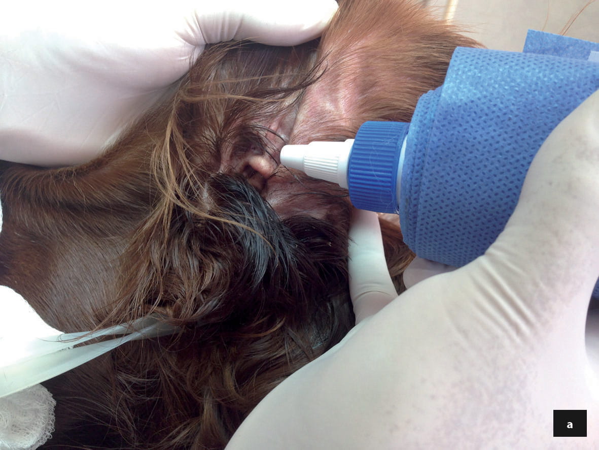
Superficial ear cleaning does not require anesthesia or sedation in the majority of patients, and owners should be instructed as to how to perform this procedure properly at home. In most cases of otitis externa, epithelial migration, the self-cleaning mechanism of the ear canal, is affected adversely, leading to cerumen accumulation 6,7.
 |
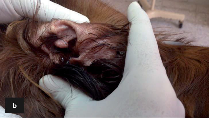 |
Ear cleaner should be installed into the ear canal (a) and the ear massaged externally (b). The cerumen may be removed from the external part of the ear using a cotton swab, but excessive use of swabs inside the ear canal should be avoided. Cleaning helps reduce the amount of cerumen exudate and facilitates the penetration of topical treatments; it also decreases the bacterial and yeast biofilm, which assists in elimination of infectious agents.
© Alberto Martín Cordero
 |
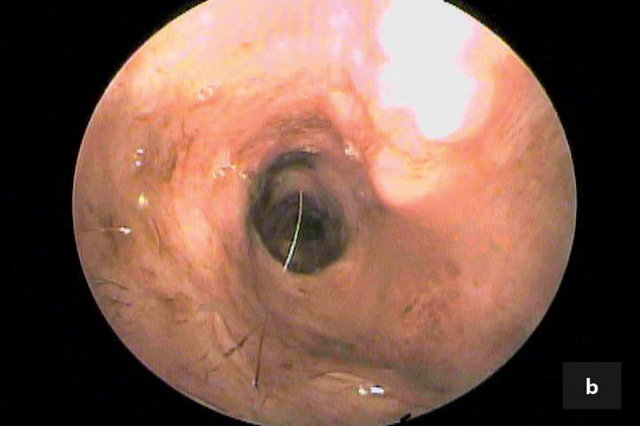 |
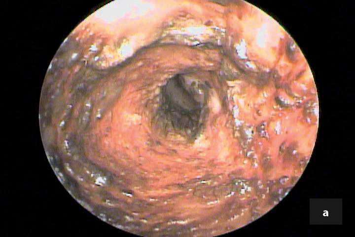
An otoscopic view of the external ear canal pre- (a) and post- (b) cleaning. In the consultation room reducing or eliminating the ceruminous debris is important, as it allows a full examination to be performed; the structures of the ear, such as the epithelium of the external canal, can be evaluated, and the tympanic membrane integrity assessed. A correct balance between treatment and control of ceruminous debris is the main goal; excessive use of cleaning agents may damage the ear canal epithelium; this may be seen as white ceruminous debris and inflammatory cells with no micro-organisms detected on cytology.
© Alberto Martín Cordero
Baba E, Fukata T, Saito M. Incidence of otitis externa in dogs and cats in Japan. Vet. Rec. 1981;108:393-395.
Griffin CE, Song M. Otitis workshop. In: Kwochka K, Willemse T, von Tscharner C (eds). Advances in Veterinary Dermatology, vol. 3. Boston: Butterworth-Heinemann 1996;369-375.
Rosychuk RA, Luttgen P. Diseases of the ear. In: Ettinger SJ, Feldman EC (eds.) Textbook of Veterinary Internal Medicine: Diseases of the Dog and Cat. 5th ed. Philadelphia: WB Saunders 2000;1185-1235.
Saridomichelakis MN, Farmaki R, Leonidas LS, et al. Aetiology of canine otitis externa: a retrospective study of 100 cases. Vet. Dermatol. 2007;18:341-347.
Campbell JJ, Coyner KS, Rankin SC, et al. Evaluation of fungal flora in normal and diseased canine ears. Vet. Dermatol. 2010;21(6);619-625.
Tabacca NE, Cole LK, Hillier A, et al. Epithelial migration on the canine tympanic membrane. Vet. Dermatol. 2011;22(6);502-510.
Nuttall T, Cole LK. Ear cleaning: the UK and US perspective. Vet. Dermatol. 2004;15(2):127-136.
Alberto Martín Cordero
Alberto Cordero is a dermatologist and founder of “Vetderm”, a referral clinic in Guadalajara, Mexico Read more
Anal and perianal pruritus can be a real nuisance and requires careful diagnosis as there are several etiologies.
Before the emergence of meticillin resistance, Staphylococcus pseudintermedius was susceptible to most of the antibiotic drugs available for animals
Immune-mediated dermatoses are uncommon diseases in the dog and cat and may be subdivided into autoimmune and immune-mediated categories
Malassezia, a genus of fungi, is frequently found as a commensal organism in the skin, ear canals, nose, oral surfaces, perianal surfaces, anal sacs, and vagina of normal dogs and cats, and has even been identified on the epidermis of puppies as young as three days old.