Worldwide medical and scientific journal for animal health professionals
Veterinary Focus

Issue number 25.2 Other Scientific

Canine cutaneous autoimmune disease

Published 01/02/2023

Written by Amy Shumaker

Also available in Français , Deutsch , Italiano and Español

Immune-mediated dermatoses are uncommon diseases in the dog and cat and may be subdivided into autoimmune and immune-mediated categories.

A mild form of discoid lupus erythematosus

Key points

Autoimmune diseases result from failure of the immune system to recognize “self”.


There are numerous autoimmune and immune-mediated cutaneous diseases that have variable clinical presentations; they can often mimic other, more common, cutaneous disorders.


Histopathologic evaluation is the gold standard in diagnosing a cutaneous autoimmune disease, but location and stage of the lesion can influence the diagnostic outcome.


Use of immunomodulatory medications versus immunosuppressive medications is dependent on the type and severity of disease.


Introduction

Immune-mediated dermatoses are uncommon diseases in the dog and cat and may be subdivided into autoimmune and immune-mediated categories 1. Autoimmunity is considered to result from failure of the immune system to recognize “self”, mounting an immune response composed of antibodies or activated lymphocytes against normal body structures and tissues, whereas immune-mediated conditions are triggered by a foreign antigen such as drugs (including vaccines) or infectious agents.

There are numerous autoimmune and immune-mediated dermatoses, with the prognosis dependent on the type of disease. Some disorders affect only the skin and have minimal or mild systemic involvement; other diseases, such as lupus erythematosus and various forms of vasculitis, can affect other organ systems and have a serious systemic impact.

This article will focus on recognition of clinical signs, diagnostic options, therapeutic modalities, and avoidance of potential triggers for autoimmune dermatoses. With the proper approach, many of these diseases can be rewarding to treat.

Clinical signs and diagnosis

As with any skin disease, diagnosis is made utilizing a combination of history, clinical signs and routine dermatologic diagnostics, such as skin scraping, cytologic analysis, and biopsy with histopathology. It is not uncommon for some disorders, such as pemphigus, to have a waxing and waning history. Most autoimmune disorders occur in young to middle-aged animals, and many auto-immune dermatoses show a breed predisposition which can aid in formulating a differential diagnosis.

Clinical presentation may be variable and can mimic many other dermatoses due to the limited number of reaction patterns of the skin. With the wide diversity of cutaneous autoimmune dermatoses, there are multiple clinical signs; whilst there is no singular “pathognomonic” sign that conveys a cutaneous autoimmune disease, the clinician may identify alopecia, crusting (e.g., pemphigus foliaceus), erythema and purpura (e.g., vasculitis, erythema multiforme), ulcerations (e.g., vasculitis, lupus/lupoid variants), and vesicles (e.g., bullous skin diseases).

The gold standard for diagnosis of autoimmune dermatoses is biopsy with histopathologic evaluation by a dermatopathologist. Multiple punch biopsies should be obtained from representative lesions; if present, areas of crusting and pustules should be biopsied for examination. Additionally, individual crusts can be submitted for evaluation for such diseases such as pemphigus. Selected sites should not be clipped or scrubbed as this can remove crusts, possibly adversely affecting results. Ideally, animals should not be on corticosteroids when biopsied, and submission of only ulcerated tissue is discouraged, as the outcome may result in an obscure diagnosis of “ulcerative dermatitis”. Special stains, including Periodic acid-Schiff (PAS), may be useful in evaluating for other mimickers, such as dermatophytosis.

Additional diagnostic considerations include cytology, dermatophyte culture, antinuclear antibody test (ANA), and tick titers. Cytology is invaluable in supporting or refuting a diagnosis of an autoimmune disease; e.g., the presence of acantholytic keratinocytes surrounded by neutrophils is highly suggestive of pemphigus foliaceus (Figure 1). However, staphylococcal infections and dermatophytes, especially Trichophyton spp., can also induce acantholysis 2. It is therefore important to evaluate for such agents and treat appropriately, if present. If bacteria are present, a 4-6-week course of systemic antibiotics should be instituted, and if resolution is noted, then a diagnosis of mucocutaneous pyoderma is supported. Note that for discoid lupus erythematosus, the clinical signs and histopathologic changes can very closely resemble mucocutaneous pyoderma of the nasal planum 3. ANA titers, as well as histopathologic analysis, can be helpful in supporting the diagnosis of systemic lupus erythematosus. Additional tests include immunofluorescence or immunohistochemical testing; direct immunofluorescence and immunohistochemical testing (often limited to specialist veterinary immunopathology laboratories) frequently requires special tissue handling, whilst indirect immunofluorescence testing on serum to detect the presence of circulating autoantibodies has shown more promise recently 1,4,5.

Impression cytology

Figure 1. Impression cytology obtained below a crust on the nasal planum of a dog diagnosed with pemphigus foliaceus. Note the number of neutrophils surrounding clusters of large, basophilic acantholytic keratinocytes (arrow) (100x magnification). 
© Amy Shumaker

Treatment

With autoimmune/immune-mediated dermatoses, there are two therapeutic approaches that can be utilized for treatment: immunosuppression or immunomodulation (Table 1a, b and c). Disease type and severity determines the approach. Most dogs with discoid lupus erythematosus, rabies vaccine-induced cutaneous vasculitis, pinnal margin vasculitis, and symmetric lupoid onychodystrophy will respond favorably to, and can be maintained on, immunomodulatory drugs. Other diseases such as pemphigus foliaceus, erythema multiforme, systemic lupus, and various other vasculitides will need immunosuppressive therapies.

Table 1a. Commonly used drugs for the treatment of cutaneous autoimmune and immune-mediated diseases. 1,24,25

Immunosuppressant drugs
Drug Dosage and notes Mechanism of action Side effects
Glucocorticoids
Prednisone/prednisolone 2.2-4.4 mg/kg q24H Dexamethasone 0.2-0.4 mg/kg q24H Triamcinolone 0.2-0.6 mg/kg q24H 
The above are induction dosages that are then tapered to the lowest dose q48H (prednisone) – 72H (dexamethasone, triamcinolone) to maintain remission
Decreases circulating levels of T-lymphocytes; inhibits lymphokines; inhibits neutrophil, macrophage and monocyte migration; inhibits phagocytosis and chemotaxis; reduces production of interferon Symptoms of hyperadrenocorticism, panting, vomiting, diarrhea, hepatic enzyme elevations, pancreatitis, GI ulceration, lipidemias, urinary tract infections, diabetes mellitus, muscle atrophy, behavioral changes
Cyclosporine
Induction: 5-10 mg/kg q24H 
Maintenance: 5-10 mg/kg q48H or less
Immunosuppressant: blocks IL-2 transcription and T-cell responsiveness to IL-2; inhibits IFN-α transcription, inhibits mononuclear cell function Vomiting, diarrhea, anorexia, gingival hyperplasia, papillomatosis, hirsutism, bacteriuria, bone marrow suppression, nephropathy
Azathioprine
Induction: 1.5-2.5 mg/kg q24H 
Maintenance: 1.5-2.5 mg/kg q48H but can be tapered to as low as 1 mg/kg q72H
Affects rapidly proliferating cells Greatest effects on cell-mediated immunity and T-cell-dependent antibody synthesis Anemia, leukopenia, thrombocytopenia, vomiting, hypersensitivity reactions, pancreatitis, elevated ALP and ALT, rashes, alopecia, diarrhea, hepatotoxicity, increased risk of infections
Mycophenolate mofetil 10-20 mg/kg q12H Inhibits de novo purine synthesis and suppresses T and B lymphocytes and production of antibodies Nausea, vomiting, diarrhea, bone marrow suppression, increased incidence of infections
Chlorambucil
Induction: 0.1-0.2 mg/kg q24-48H 
Maintenance: 0.1-0.2 mg/kg q48H or less
Cytotoxic effects via cross-linking of DNA Anorexia, vomiting, diarrhea, myelosuppression
Cyclophosphamide
1.5 mg/kg q48H 
Due to side effects, often recommended for use in induction phase only; rarely used currently for cutaneous autoimmune diseases
Inhibits mitosis; immunosuppressive to humoral and cell-mediated systems, suppresses antibody production Sterile hemorrhagic cystitis, bladder fibrosis, teratogenesis, infertility, alopecia, nausea, GI inflammation, increased infections, bone marrow suppression


Immunomodulatory drugs may take time to effect an improvement (generally seen within 3-4 weeks of starting therapy) so if the clinical signs are severe, a tapering course of high dose glucocorticoids can be utilized initially to obtain rapid control, along with a chosen immunomodulatory drug. Once remission is achieved the immunomodulatory drug can be continued as maintenance. Note that both the glucocorticoids and the immunomodulatory drug should be given initially since the latter class of drug can take time to be effective: this will help prevent relapse of the disease once steroids are tapered. The primary benefit of immunomodulatory drugs is that they have less serious adverse side effects and decreased health impact.

When immunosuppressive therapy is utilized, the most commonly used drug is a glucocorticoid. Initially, high doses are needed to achieve remission, and then tapered to the lowest possible dose that will maintain remission with minimal adverse systemic effects. In many autoimmune diseases, adjunctive therapies are necessary in order to permit the glucocorticoid dose to be lowered to a level which minimizes adverse side effects. In the more severe cases, it is not unusual to combine several different immunosuppressant drugs to achieve and maintain remission. As many of these medications can have adverse side effects on the liver and bone marrow, blood monitoring every 2-3 weeks for the first several months is recommended, with maintenance monitoring every 4-6 months. If significant changes in the blood parameters are noted, the offending drug should be discontinued and replaced with another medication. The most commonly utilized adjunctive medications include azathioprine, cyclosporine, mycophenolate mofetil, cyclophosphamide, and chlorambucil. In more severely affected dogs, supportive care for open wounds, fluid corrective therapy and monitoring of serum protein levels may be necessary. Use of human intravenous immunoglobulin (hIVIg) has shown promise in treating severe autoimmune dermatoses when other treatments are failing 6.

Topical therapies can be useful with more localized lesions or for sporadic flare-ups. The most commonly used topicals include betamethasone or tacrolimus. Betamethasone has the benefit of rapidly controlling inflammation and disease symptoms but can induce dermal atrophy with chronic use; therefore, transition to tacrolimus is prudent if a topical is needed for long-term use.

Table 1b. Commonly used drugs for the treatment of cutaneous autoimmune and immune-mediated diseases. 1,24,25

Immunomodulatory drugs
Drug Dosage and notes Mechanism of action Side effects
Tetracyclines
Doxycycline: 5 mg/kg q12H 
Minocycline: 5-10 mg/kg q12H Tetracycline: 
500 mg for dogs >10 kg q8H 
250 mg for dogs <10 kg q8H
Anti-inflammatory properties that affects chemotaxis, antibody production, complement activation; down-regulates cytokines; inhibits prostaglandin synthesis, lipases and collagenases Vomiting, anorexia, lethargy, diarrhea, increased liver enzyme activity
Niacinamide
500 mg for dogs > 10 kg q12H* 
250 mg for dogs < 10 kg q12H* 
*Given q8H if administered with tetracycline
Blocks antigen IgE-induced histamine release and degranulation of mast cells; photoprotectant and cytoprotectant that blocks inflammatory cell activation and apoptosis; inhibits phosphodiesterases; decreases protease release Anorexia, vomiting, lethargy, occasional hepatic enzyme elevations
Pentoxifylline
10-30 mg/kg q8-12H
Inhibits erythrocyte phosphodiesterase and decreases blood viscosity, increasing erythrocyte flexibility, reduces negative endotoxic effects of cytokine mediators Vomiting, anorexia, CNS excitement or nervousness


There are four phases to consider in the treatment of cutaneous autoimmune dermatoses: induction phase, transition phase, maintenance phase and determining cure 1. With the induction phase, the goal is to stop the inflammatory component as quickly as possible and suppress the immunologic response directed towards the skin. In this phase, higher doses of medications are normally necessary. If an acceptable response is not noted in a timely manner, another treatment regimen should be considered; i.e., alternative medications chosen or additional medications added to the current treatment regimen. In the transition phase, drugs are tapered to minimize side effects and adverse reactions. When drug combinations are utilized, those with the greatest side effects – such as glucocorticoids – are the first to be tapered. Medications are slowly reduced, often over several weeks or months, until an acceptable maintenance dose of medications is achieved, or until signs recur. If this happens, the medications are increased until remission is again achieved, then tapered down to the last dose that maintained the patient’s symptoms under acceptable control (the maintenance phase). A “cure” is considered for cases of immune-mediated dermatoses that have achieved remission and are successfully controlled with maintenance therapy but do not then recur after cessation.

Cessation of maintenance therapy in a patient that has been well-controlled is a difficult decision to make, especially if the initial disease was severe. This decision should be one that is mutually agreed between the clinician and owner; it is essential that the client is well-informed, with the realization that if the patient relapses, achieving remission a second time may be more difficult. When to discontinue maintenance therapy depends on the type of disease, whether or not a trigger was identified and removed, and the risk to the patient if therapy is discontinued. In many cases, maintenance therapy for 8-12 months is recommended before cessation 1. In animals where the risk of recurrence outweighs the benefit of discontinuing therapy, the drugs can be maintained life-long with appropriate lab work monitoring.

Future vaccinations are often discouraged in cases of autoimmune dermatoses, even in those where vaccination is not a known trigger. Concern lies with the idea that vaccination may stimulate a broad, non-specific immune response, possibly initiating recrudescence of the autoimmune disease 7. The author prefers to discontinue rabies vaccination and monitor titer levels for distemper and parvovirus. If titers are not sufficient to maintain proper immunity, a risk vs. benefit assessment should be performed before considering re-vaccination.

Table 1c. Commonly used drugs for the treatment of cutaneous autoimmune and immune-mediated diseases. 1,24,25

Topicals
Drug Dosage and notes Mechanism of action Side effects
Tacrolimus 0.1%
Applied 1-2 times per day then taper down to less frequent use
Inhibits T-cell activation and proliferation via cytokine suppression Localized erythema, irritation, pruritus Owners should wear gloves
Betamethasone 0.1%
Applied 1-2 times per day then taper down to less frequent use (ideally twice weekly for chronic use)
Similar effects as with systemic glucocorticoids; inhibit migration of lymphocytes and macrophages locally Dermal atrophy; increased risk of induction of hypothalamic-pituitary-adrenal axis suppression; systemic glucocorticoid effects; development of milia and comedones; local skin reactions


Specific diseases

Pemphigus foliaceus

The most common autoimmune skin disorder in the dog, pemphigus foliaceus (PF) is a pustular to crusting autoimmune dermatitis. PF affects the epidermis, targeting various adhesion molecules, especially desmosomes, which hold keratinocytes together. In human PF, the desmoglein-1 glycoprotein (DSG1) in the desmosome is the primary target of autoantibodies 8 and the same glycoprotein was previously suspected to be the primary target in dogs 9,10; however, it is now believed to be a minor autoantigen 11, with current evidence suggesting that desmocollin-1 is a major autoantigen in canine PF 12.

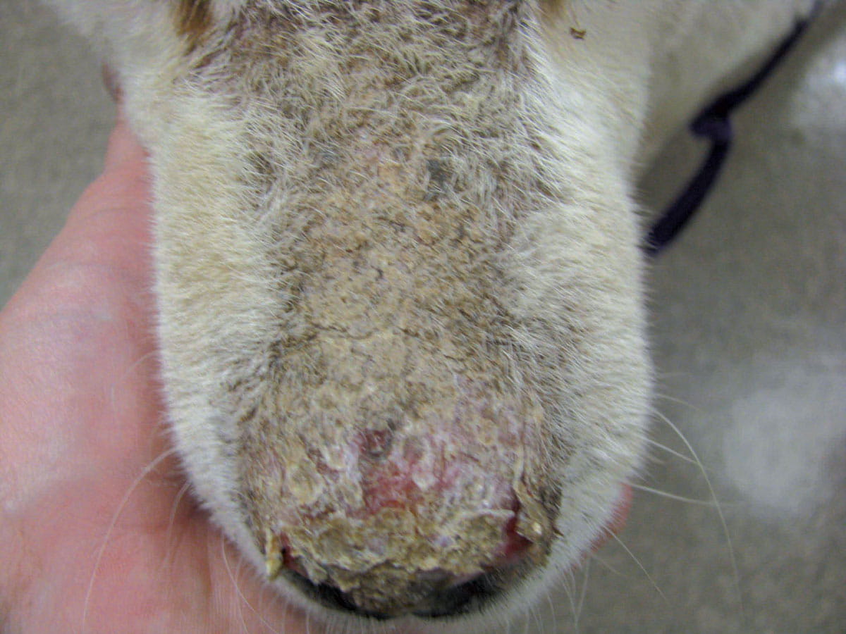
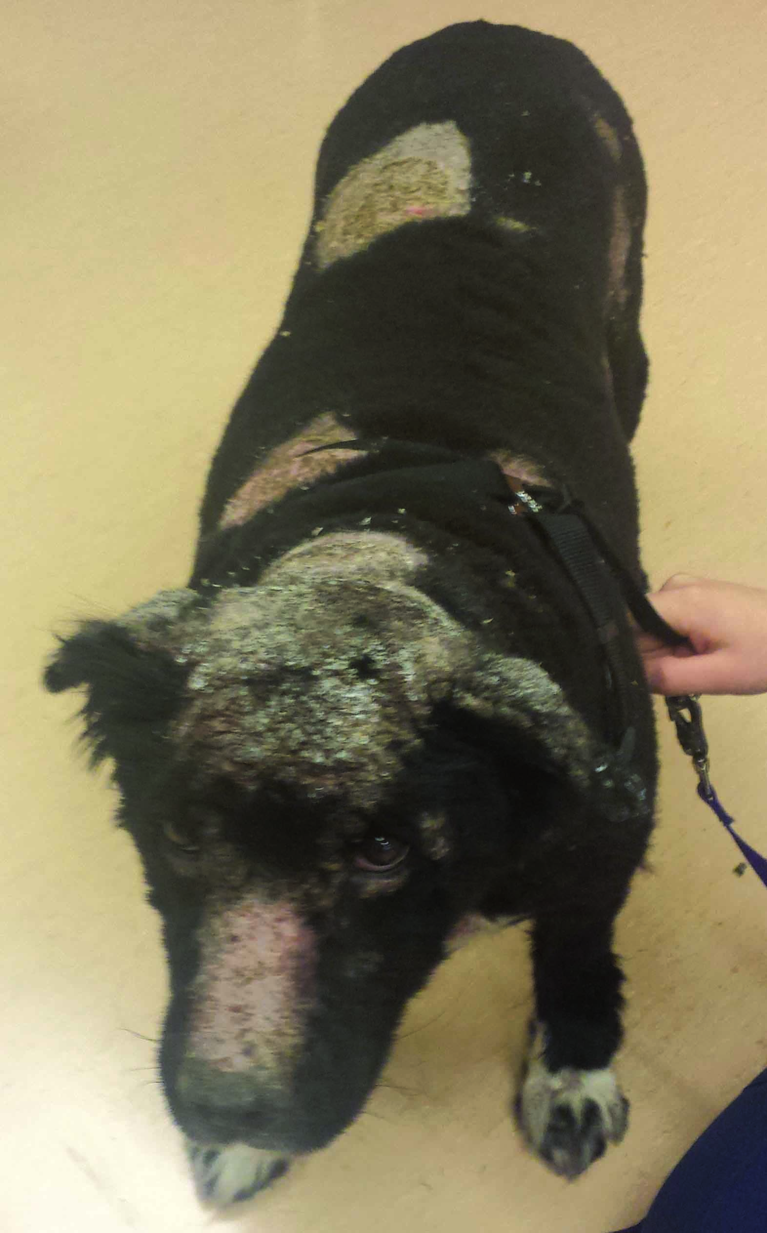
Genetic factors appear to play a role in the development of PF, with Akitas and Chow Chows the breeds considered most at risk 10. Triggers include chronic allergic skin disease and drugs (antibiotics, NSAIDs, topical flea spot-ons), but the most important one is ultraviolet light 1,10. The initial lesion is a macule that rapidly progresses to pustules, which are often large and coalesce. The pustules are frequently fragile and easily ruptured, resulting in crusting. As a result, crusts are the most common clinical sign 1,9,10. Erosions may be noted; ulcerations are rare but can be present in cases complicated with a deep pyoderma. Canine PF is often characterized by crusting, initially involving the face (especially dorsal muzzle and nasal planum, peri-ocular region and pinnae), and subsequently progressing to a generalized form (Figure 2).

Cytology of an intact pustule or of the skin below a crust will often reveal the presence of numerous non-degenerate neutrophils surrounding individual or rafts of acantholytic keratinocytes, which appear as large, rounded basophilic nucleated keratinocytes (Figure 1). Histological evaluation reveals subcorneal pustules containing neutrophils and variable numbers of eosinophils, and acantholytic keratinocytes 13. Treatment often involves high doses of steroids with an adjunctive immunosuppressant and topicals for localized treatment.

Honey-colored crusting involving the dorsal muzzle and nasal planum  
a
 A more generalized presentation of pemphigus foliaceus
b


Figure 2. Pemphigus foliaceus: (a) Honey-colored crusting involving the dorsal muzzle and nasal planum is depicted. Mild erosion of the nasal planum can be seen underlying the lifted crust. (b) A more generalized presentation of pemphigus foliaceus. 
© Amy Shumaker


Discoid lupus erythematosus

Also referred to as “collie nose” or cutaneous lupus erythematosus, discoid lupus erythematosus (DLE) is a benign ulcerative disease without systemic manifestations 1. DLE is generally localized to the nasal planum, but can involve the sun-exposed areas of the pinnae and peri-ocular region and there are reports of generalized variants 14. The most common clinical sign is initial loss of the cobblestone architecture of the nasal planum progressing to depigmentation and scaling (Figure 3). With chronicity, erosions, ulcerations and crusting occur. Annular to polycyclic hyperpigmented plaques involving the neck, trunk and extremities may be seen in the generalized variant cases.

Histopathology reveals interface basal cell degeneration (apoptosis), with a moderate pleocellular lichenoid infiltrate of the dermis 13. As this disease can closely mimic mucocutaneous pyoderma both clinically and histopathologically, cytologic evaluation of the nasal planum below a crust can be useful; treatment of mucocutaneous pyoderma is recommended if bacteria are present.

In most cases of DLE, the use of potent immunosuppressants are unnecessary; a systemic immunomodulatory approach utilizing a tetracycline (doxycycline, minocycline) and niacinamide coupled with topical therapy (topical steroids, tacrolimus) is often successful at controlling the disease. In refractory or severe cases, high doses of corticosteroids may be needed initially. In the reported generalized variants, hydroxychloroquine or cyclosporine were effective treatments 14,15. As sunlight plays a significant role in DLE, it is important that sun exposure be minimized with avoidance and use of sunscreens. Vitamin E supplementation (400 IU daily) may also be helpful.

discoid lupus erythematosus

Figure 3. A mild form of discoid lupus erythematosus in a dog with chronic sun exposure; note the loss of cobblestone architecture of the nasal planum with depigmentation and focal erosions. 
© Amy Shumaker

Erythema multiforme

A rare immune-mediated dermatosis, erythema multiforme (EM) can be idiopathic in nature or triggered by numerous factors, including drugs, bacterial infections, parvovirus, food, vaccination, and neoplasia 1,16,17. In one review of 44 dogs with EM, drugs were the trigger in 26 (59%) cases 16; the most commonly implicated drugs are antibiotics such as trimethoprim-potentiated sulfonamides, penicillins and cephalosporins. EM has been subclassified into major and minor forms. EM minor is mild, with acute onset of the typical target lesions most often involving the extremities, with no or slight mucosal involvement; if present, it is limited to the oral mucosa, and systemic symptoms are not noted. EM major is more severe, with significant mucosal involvement and often constitutional symptoms such as lethargy and pyrexia. The distinction between EM major and Stevens-Johnson syndrome (SJS) can be difficult, and it is possible that many cases diagnosed as EM may actually be a result of SJS 1. Skin lesions are variable (Figure 4), and this disease can mimic many other dermatoses; however, lesions may have an acute onset and are often symmetrical, consisting of erythematous macules or elevated papules that spread peripherally, clearing centrally. Many can have an annular to arciform or serpiginous pattern. Additional lesions include urticarial plaques, and vesicles and bullae that progress to ulcers. Mucosal lesions are generally erythematous and can also progress to vesicular, bullous and ulcerative lesions; crusting may be associated with some lesions. The most commonly affected sites include the ventrum, axillae, mucocutaneous junctions, oral cavity, pinnae and footpads.

With such variation in clinical signs and a large differential diagnosis to consider based on clinical signs (bacterial folliculitis, demodicosis, dermatophytosis, urticaria, other vesicular and bullous disorders), biopsy with histopathology is needed for diagnosis. The most characteristic histopathologic feature with EM is panepidermal apoptosis with lymphocyte satellitosis and an interface dermatitis 13. Response to treatment and perhaps permanent remission is contingent upon identifying and eliminating a trigger where one can be found, as elimination of the etiology can result in spontaneous resolution within weeks of correcting and treating. A hypo-allergenic elimination diet trial should be performed in cases with no identifiable trigger, as food hypersensitivity can be a potential cause 18. In more severe cases, and for those cases where a trigger cannot be identified, immunosuppressants such as corticosteroids, azathioprine, and cyclosporine have been effective. In life-threatening cases, hIVIg has been utilized to improve and expedite treatment outcome 1,19.

Erythema multiforme

Figure 4. Erythema multiforme exhibiting various clinical lesions, including patchy alopecia, scaling, erythema, erosions and ulcerations. Note the lesional changes on the eyelids consistent with the mucocutaneous involvement found with this condition. 
© Katherine Doerr, DVM, DACVD

Cutaneous vasculitis

There are a variety of vascular diseases that affect dogs. Cutaneous vasculitis is a disease process whereby blood vessel walls are targeted by an inflammatory response, resulting in subsequent destruction of blood vessels and ischemic necrosis of the affected tissue. It is important to note that cutaneous vasculitis is more a cutaneous reaction pattern rather than a specific diagnosis, as there are multiple causes that trigger vasculitis. Cutaneous vasculitis has been associated with other co-existing diseases, including food hypersensitivity, insect bites, malignancies, and infectious diseases including tick-borne diseases 20,21,22. Additionally, numerous drugs have been implicated as causing vasculitis 21,22,23. In many cases, an underlying etiology is not readily identifiable, and the disease is considered idiopathic. In most cutaneous vasculitides, the pathomechanism is suspected to be a type III hypersensitivity reaction, whereby immune complexes formed following antigen exposure are deposited in vessel walls. However, additional factors may be involved, including genetics, defects in immune complex clearance, and autoantibodies.

The skin may be the only organ involved with vasculitis, but other organs may also be affected, such as the kidneys in Greyhounds. Typical cutaneous lesions of cutaneous vasculitis include palpable purpura, erythemic to purpuric plaques, hemorrhagic bullae, with progression of the disease resulting in development of demarcated ulcers involving paws, pinnal apex, lips, tail and oral mucosa 20. Pitting edema may also be present. In some cases, the claws may be affected and exhibit signs of onychodystrophy, onychomadesis, petechiae, and exudate within the claw. Erosive, ulcerative or hyperkeratotic lesions may affect the pads. Often, the ulcerations or depressions will affect the center of the pad; however, lateral margins may also be affected. In rabies vaccine-induced vasculitis, an annular patch of alopecia with variable degrees of hyperpigmentation (Figure 5), erythema and occasional scaling occurs at the site of vaccination, usually within 2-6 months of vaccination. In these cases, additional areas may be affected, especially the apex of the pinnae.

An alopecic, hyperpigmented patch

Figure 5. An alopecic, hyperpigmented patch consistent with ischemic dermatopathy following rabies vaccination. 
© Amy Shumaker

Diagnosis is made by histopathologic evaluation; however, changes can often be subtle depending on the stage of the disease and the site selected for biopsy, making diagnosis difficult. Histological findings typical of vasculitis include variable degrees of neutrophilic, eosinophilic and mononuclear cell invasion of vessel walls with endothelial cell swelling, fibrinoid degeneration, red blood cell extravasation, and occasional leukocytoclasia noted within or near the vessel walls 13,20.

Other dermal changes include pale-staining collagen, follicular atrophy, and cell-poor interface dermatitis 1,13. In cases of vaccine-induced vasculitis, an amorphous basophilic material may be noted representing likely vaccine product 13. The type of cellular inflammation present may be indicative of the trigger; for example, an eosinophilic vasculitis is often associated with arthropod reactions, food hypersensitivity, mast cell tumors or canine eosinophilic dermatitis.

When a diagnosis of vasculitis is made, additional work up may be needed to determine the underlying cause. A thorough history should be taken and any recent drug or vaccine administration should be investigated. Tick titers should be performed. An elimination diet trial with a commercial novel protein or hydrolyzed protein diet may be useful if food hypersensitivity is suspected, especially in cases of urticarial vasculitis.

Treatment of vasculitis is dependent on the severity of disease and type of vasculitis. Treatment length is also variable, as some cases may resolve and go into permanent remission if an underlying trigger can be identified and eliminated. Other cases may require life-long therapy. In more severe cases, treatment with glucocorticoids (with or without an adjunctive immunosuppressant drug) may be necessary once infectious causes have been ruled out. In cases of vaccine-induced vasculitis, immunomodulatory therapy utilizing a combination of medications, including doxycycline/minocycline, niacinamide and pentoxifylline, is often successful. Topical therapies containing steroids, such as betamethasone, can be utilized short term for more localized lesions, with transition to tacrolimus if longer treatment is necessary for topical control.

Conclusion

Cutaneous autoimmune and immune-mediated diseases are uncommon to rare in dogs, but are still likely to be encountered in general practice. As many disorders can mimic cutaneous autoimmune disease – and vice versa – a thorough history and diagnostic work-up is imperative to achieve an appropriate diagnosis and treatment regimen, with identifiable triggers eliminated. Where appropriate, immunomodulatory therapy, rather than immunosuppressant therapy, should be considered as there are perceived less systemic side effects, but life-long therapy may be necessary in many cases.

References

  1. Miller WH, Griffin CE and Campbell KL. Autoimmune and immune-mediated dermatoses. In: Muller and Kirk’s Small Animal Dermatology 7th Ed. St. Louis, MO: Saunders, 2013;439-500. 

  2. Olivry T, Linder K. Dermatoses affecting desmosomes in animals: a mechanistic review of acantholytic blistering diseases. Vet. Dermatol. 2009;20:313-326. 

  3. Wiemelt SP, Goldschmidt S, Greek JS, et al. A retrospective study comparing the histopathological features and response to treatment in two canine nasal dermatoses, DLE and MCP. Vet. Dermatol. 2004;15:341-348. 

  4. Nishifui K, Tamura K, Konno H, et al. Development of an enzyme-linked immunosorbent assay for detection of circulating IgG autoantibodies against canine desmoglein-3 in dogs with pemphigus. Vet. Dermatol. 2009;20(5-6):331-337. 

  5. Bradley GA, Mays MB. Immunoperoxidase staining for the detection of autoantibodies in canine autoimmune skin disease; comparison of immunofluorescence results. Vet. Immunol. Immunopathol. 1990;26(2):105-113. 

  6. Spurlock NK, Prittie JE. A review of current indications, adverse effects, and administration recommendations for intravenous immunoglobulin. J. Vet. Emerg. Crit. Care 2011;21(5):471-483. 

  7. Westra J, Rondaan C, van Assen S, et al. Vaccination of patients with autoimmune inflammatory rheumatic diseases. Nat. Rev. Rheumatol. 2015;11(3):135-145. 

  8. Stanely JR, Koulu L, Klaus-Kovtun V, et al. A monoclonal antibody to the desmosomal glycoprotein desmoglein-1 binds the same polypeptide as human autoantibodies in pemphigus foliaceus. J. Immunol. 1986;136(4): 1227-1230. 

  9. Mueller RS, Krebs I, Power HT, et al. Pemphigus foliaceus in 91 dogs. J. Am. Anim. Hosp. Assoc. 1989;194(4):545-546. 

  10. Olivry T. A review of autoimmune skin diseases in animals: 1 – superficial pemphigus. Vet. Dermatol. 2006;17(5):291-305. 

  11. Olivry T, LaVoy A, Sunston SM, et al. Desmoglein-1 is a minor autoantigen in dogs with pemphigus foliaceus. Vet. Immunol. Immunopathol. 2006;111 (3-4):245-255. 

  12. Bizikova P, Dean GA, Hashimoto T, et al. Cloning and establishment of desmocollin-1 as a major autoantigen in canine pemphigus foliaceus. Vet. Immunol. Immunopathol. 2012;149(3-4):197-207. 

  13. Gross TL, Ihrke PE, Walder EJ, et al. Skin diseases of the dog and cat, 2nd ed. Ames, Iowa: Blackwell, 2005;65-68, 247-248, 263-267, 415-416. 

  14. Oberkirchner U, Linder KE, Olivry T. Successful treatment of a novel generalized variant of canine discoid lupus erythematosus with oral hydroxychloroquine. Vet. Dermatol. 2012;23(1):65-70. 

  15. Benovic F, Olivry T, Linder KE. Cyclosporin therapy for canine discoid lupus erythematosus refractory to doxycycline and niacinamide. Vet. Dermatol. 2014;25(5):483-e79. 

  16. Scott DW, Miller WH. Erythema multiforme in dogs and cats: Literature review and case material from the Cornell University College of Veterinary Medicine (1988-1996). Vet. Dermatol. 1999;10:297-309. 

  17. Kang MH, Park HM. Erythema multiforme minor in a dog following inappropriate intranasal Bordetella bronchiseptica vaccination: a case report. Veterinarni Medicina 2011;56(11):568-572. 

  18. Itoh T, Nibe K, Kojimoto A, et al. Erythema multiforme possibly triggered by a food substance in a dog. J. Vet. Med. Sci. 2006;68(8):869-871. 

  19. Nuttal T, Malham T. Successful intravenous human immunoglobulin treatment of a drug-induced Stevens-Johnson syndrome in a dog. J. Small Anim. Pract. 2004;45(7):357-361. 

  20. Nichols PR, Morris DO, Beale KM. A retrospective study of canine and feline cutaneous vasculitis. Vet. Dermatol. 2001;12(5):255-264. 

  21. Innera M. Cutaneous vasculitis in small animals. Vet. Clin. North Am. Small Anim. Pract. 2013;43(1):113-134. 

  22. Nichols PR, Morris DO, Beale, KM. A retrospective study of canine and feline cutaneous vasculitis. Vet. Dermatol. 2001;12(5):255-264. 

  23. Niza MM, Felix N, Vilela CL, et al. Cutaneous and ocular adverse reactions in a dog following meloxicam administration. Vet. Dermatol. 2007;18(1):45-49. 

  24. Rosenkrantz W. Pemphigus: current therapy. Vet. Dermatol. 2004;15(2):90-98. 

  25. Plumb D. Plumb’s Veterinary Drug Handbook, 7th ed. Ames, Iowa: Wiley Blackwell, 2001. 90-102, 110-112, 195-197, 259-262, 262-266, 471-472, 698-699, 720-721, 735-736, 801-803, 967-969, 1132-1133.

Amy Shumaker

Amy Shumaker

Dr. Shumaker graduated from the University of Florida, College of Veterinary Medicine in 2001 Read more

Other articles in this issue

Issue number 25.2 Published 03/05/2023

Ear infection: what the owner needs to know

Otitis externa can be frustrating for both the owner and the clinician, as treatment requires a lot of effort, often for a protracted period of time. This article details the minimum information that should be provided to the cat and dog owner when the problem is first identified.

By Alberto Martín Cordero

Issue number 25.2 Published 05/04/2023

Perianal pruritus in the dog

Anal and perianal pruritus can be a real nuisance and requires careful diagnosis as there are several etiologies.

By Elisa Maina and Chiara Noli

Issue number 25.2 Published 22/03/2023

Canine pyoderma: the problem of meticillin resistance

Before the emergence of meticillin resistance, Staphylococcus pseudintermedius was susceptible to most of the antibiotic drugs available for animals

By Ana Oliveira

Issue number 25.2 Published 18/01/2023

Malassezia dermatitis and otitis in dogs

Malassezia, a genus of fungi, is frequently found as a commensal organism in the skin, ear canals, nose, oral surfaces, perianal surfaces, anal sacs, and vagina of normal dogs and cats, and has even been identified on the epidermis of puppies as young as three days old.

By Katherine Doerr