Worldwide medical and scientific journal for animal health professionals
Veterinary Focus

Issue number 25.2 Other Scientific

Malassezia dermatitis and otitis in dogs

Published 18/01/2023

Written by Katherine Doerr

Also available in Français , Deutsch , Italiano and Español

Malassezia, a genus of fungi, is frequently found as a commensal organism in the skin, ear canals, nose, oral surfaces, perianal surfaces, anal sacs, and vagina of normal dogs and cats, and has even been identified on the epidermis of puppies as young as three days old.

A skin biopsy

Key points

Malassezia pachydermatis is a common cause of dermatitis and otitis in dogs. 


The clinical signs are due to release of virulence factors from Malassezia organisms and the resultant inflammatory cascade within the skin.


Typical clinical signs include pruritus, erythema, scaling, waxy exudation, and lichenification.


Cytological examination is the most beneficial and convenient method for diagnosis of Malassezia dermatitis.


Treatment must be focused on resolving the underlying cause of the Malassezia dermatitis; topical therapy is the mainstay for both treatment and management, while systemic therapy may be utilized in severe or refractory cases.


Introduction

Malassezia, a genus of fungi, is frequently found as a commensal organism in the skin, ear canals, nose, oral surfaces, perianal surfaces, anal sacs, and vagina of normal dogs and cats 1,2,3,4, and has even been identified on the epidermis of puppies as young as three days old 5. However, it can also be involved with dermatological disease; common clinical manifestations include hyperpigmentation, seborrhea oleosa, erythema, and variable degrees of pruritus (Figure 1).

The most common species of yeast isolated in the dog is Malassezia pachydermatis (also known as Pityrosporum canis, Pityrosporum pachydermatis, and Malassezia canis). This non-mycelial organism is a non-lipid-dependent, lipophilic, saprophytic yeast that reproduces asexually by sympodial or monopolar budding. Malassezia obtusa, M. restricta, M. sloofiae, M. furfur (also known as Pityrosporum ovale), and M. sympodialis are all lipid-dependent lipophilic species that have also been isolated from the skin and ears of dogs and cats, but less commonly 6.

Malassezia pachydermatis has significant genetic diversity; seven sequevars or strains (1a through 1g) of the organism have been identified 7; sequevar type 1a was the most prevalent and found in all host species, while type 1d was found solely in dogs. None of the sequevar types have been specifically associated with either healthy or lesional skin at this time, and more than one sequevar may colonize a single host 8,9.

A mixed-breed dog with severe lichenification, erythema, and alopecia

Figure 1. A mixed-breed dog with severe lichenification, erythema, and alopecia due to M. pachydermatis
© Dr. Stephen White/UC Davis VMTH

Pathogenesis

Multiple factors are involved in the pathogenesis of Malassezia dermatitis, such as the host’s corneocyte adherence mechanisms and the prevalence of concurrent symbiotic organisms, as well as the host’s immune response.

Adherence to canine corneocytes may be an important factor in the pathogenesis of Malassezia dermatitis in some dogs. Yeast cell walls, which consist of chitin, glucan, chitosan, and mannan 1, contain trypsin-sensitive proteins or glycoproteins that contribute to adherence to canine corneocytes. M. pachydermatis also specifically expresses adhesins that bind to mannosyl-bearing carbohydrate residues on the canine corneocyte. This adherence mechanism, however, does not apparently play a role in the pathogenesis of Malassezia dermatitis in Basset Hounds (which are prone to Malassezia overgrowth) but seems to be significant in other breeds 10.

M. pachydermatis appears to have a symbiotic relationship with commensal staphylococci species, although suggestions that Malassezia dermatitis is associated with prior antibiotic therapy have not been substantiated. The two organisms produce growth factors and micro-environmental alterations that are mutually beneficial; thus, there are increased numbers of Staphylococcus pseudintermedius or S. intermedius on dogs with concurrent Malassezia 1,4,8. In fact, 40% of dogs with Malassezia overgrowth are diagnosed with staphylococcal pyoderma due to the symbiotic relationship between the two organisms 3,11.

The yeast may elicit a spectrum of immunological responses in the host. The humoral response is stimulated, exemplified by higher numbers of antibodies found against more antigens in dogs with Malassezia versus healthy dogs 12,13. However, the elevated IgA and IgG levels found in dogs with Malassezia dermatitis does not appear to offer any additional protection against infection with the yeast. Cell-mediated immunity may play a larger role in protection against disease than humoral immunity. For example, Basset Hounds appear to have decreased lymphocyte responses to Malassezia versus healthy dogs which do not get overgrowth of the yeast 14.

Another type of immunological response that can occur in dogs is a hypersensitivity or inflammatory reaction. The preceding reactions to yeast products and antigens appear to be the main culprit in the pathogenesis of Malassezia dermatitis, since the yeast itself remains in the upper level of the epidermis 4,8. As the yeast adheres to the canine corneocytes, it secretes various substances including zymosan, urease, proteases, phosphohydrolase, phospholipases (especially phospholipase A2), lipoxygenases, phosphatases, glucosidase, galactosidase, and leucine arylamidase. These virulence factors cause alteration of the local pH, proteolysis, lipolysis, complement activation, and eicosanoid release in the skin, thus inciting the inflammatory response and pruritus 1,4,8. Furthermore, higher levels of Malassezia-specific IgE to allergens of 45, 52, 56 and 65 kDa have been found in atopic dogs compared to normal dogs, further substantiating the hypersensitivity potential of yeast 15.

Factors that predispose to pathogenicity

Factors that may predispose M. pachydermatis to become pathogenic, rather than remain commensal, may include any of the following: increased humidity, skin folds, endocrine diseases, keratinization disorders, genetic predisposition, immunologic dysfunction, hypersensitivity diseases, and increased numbers of symbiotic staphylococci.

Humidity may be important as Malassezia organisms appear to be more common in otic canals and skin folds and prevalence increases in humid climates 1. Endocrine diseases, such as hypothyroidism, primary and iatrogenic hyperadrenocorticism, and diabetes mellitus may allow increased availability of nutrients and growth factors for the yeast. This may be due to changes in cutaneous fatty acid concentrations, abnormal keratinocyte lipogenesis, and alterations in sebaceous gland function 16,17. American Cocker Spaniels, Shih Tzus, English Setters, West Highland White Terriers, Basset Hounds, Toy and Miniature Poodles, Boxers, Australian and Silky Terriers, Cavalier King Charles Spaniels, Dachshunds, and German Shepherd dogs appear to be at a higher risk for Malassezia dermatitis, suggesting a genetic component to the disease 4,6,8. Dysfunction in secretory IgA or cell-mediated immunity may also contribute to pathogenicity in some dogs 2,4. For example, Basset Hounds with Malassezia dermatitis have decreased in vitro lymphocyte blastogenic response to M. pachydermatis antigen when compared to healthy Bassets, indicating a cell-mediated immune dysfunction 14. Hypersensitivity diseases, such as flea allergy dermatitis, cutaneous adverse food reaction, and atopic dermatitis, may also predispose dogs to Malassezia dermatitis due to incitation of the inflammatory cascade and resultant pruritus.

In summary, any dermatoses that produces disruption of the stratum corneum barrier, whether mechanical (due to pruritus), or biochemical (due to endocrinopathies, keratinization or immunologic disorders), may potentially allow Malassezia virulence factors to gain exposure to the subcorneal immune system, resulting in pathogenicity of the yeast.

Diagnosis

Clinical presentation

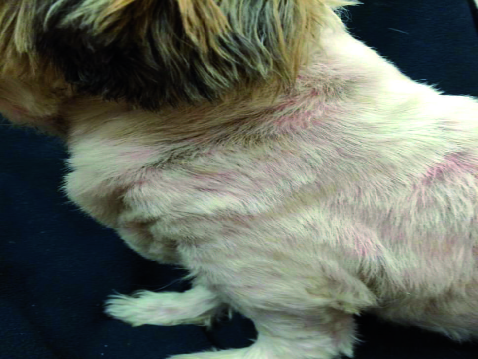
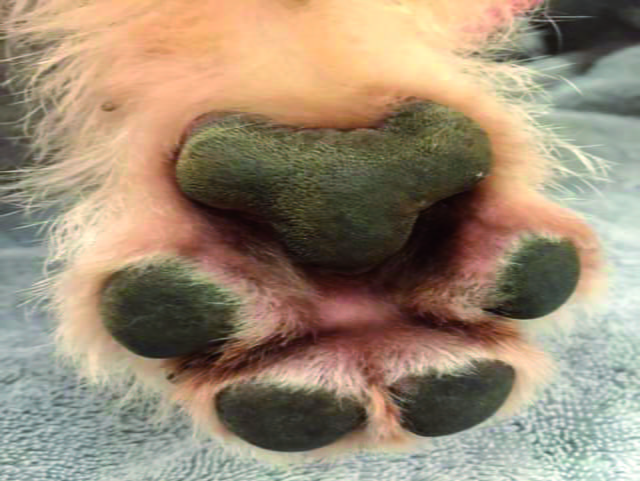
Dermatological lesions of Malassezia dermatitis may be localized (Figure 2) or generalized. They commonly manifest in warm, moist areas such as lip folds, otic canals, axillae, groin, ventral neck, medial thighs, interdigital skin, perianal and perivulvar regions, and other intertriginous areas (Figure 3). Concurrent dermatoses, such as staphylococcal pyoderma, allergies, or keratinization disorders, are seen in 70% of affected dogs 1,4. Appearance of the lesions commonly starts in the humid summer months – which is also the peak time for seasonal allergies – and may persist through the winter months. Historically, patients lack a response to glucocorticoids.

A dog with peri-ocular Malassezia dermatitis

Figure 2. A dog with peri-ocular Malassezia dermatitis. 
© Dr. Stephen White/UC Davis VMTH

A dog with Malassezia dermatitis

Figure 3. A dog with Malassezia dermatitis and accompanying lichenification, erythema and alopecia.
© Dr. Stephen White/UC Davis VMTH

A consistent clinical sign is pruritus, which can be mild to severe 1. Physical examination findings may vary, but most commonly consist of erythema (Figures 4 and 5), yellow/gray adherent or non-adherent scaling, and occasionally adherent crusting. Other manifestations include a papulocrustous dermatitis, interdigital cysts, reddish-brown discoloration of the claw beds and claws (Figure 6), erythematous macules and patches, and malodor. Secondary lesions, such as waxy or greasy exudation, lichenification, hyperpigmentation, and excoriations, may also be detected.

dog with diffuse Malassezia dermatitis
a
dog with Malassezia pododermatitis
b

Figure 4 shows a dog with diffuse Malassezia dermatitis whilst Figure 5 depicts a dog with Malassezia pododermatitis. Erythema is a common finding in dogs with Malassezia
© Dr. Katherine Doerr/Dermatology for Animals

Differential diagnoses for Malassezia dermatitis may include one or more of the following: superficial staphylococcal folliculitis, demodicosis, scabies, dermatophytosis, fleabite hypersensitivity, cutaneous adverse food reaction, contact dermatitis, atopic dermatitis, seborrheic dermatitis, epitheliotropic lymphoma, and acanthosis nigricans. It is important to rule out each differential possibility by specific diagnostic methods in order to achieve successful patient management.

A Shih Tzu dog with Malassezia paronychia

Figure 6. A Shih Tzu dog with Malassezia paronychia. 
© Dr. Katherine Doerr/Dermatology for Animals

Cytology

Cytological examination is the most beneficial and convenient method for diagnosis of Malassezia dermatitis 1. There are multiple modalities to obtain the cytological sample, including superficial skin scraping, clear acetate or cellophane tape stripping, direct impression smears, and cotton swab smears 1,4. Tape stripping can be quite effective in many anatomical locations, including dry and greasy lesions. Cotton swab impressions, while effective for otic samples, appear to be significantly inferior to direct impression, tape stripping, and superficial scraping methods in recovering yeasts from the skin 18. The sample is affixed to a glass slide, heat-fixed (if not tape), and stained with a commercial Romanowsky-type stain. Tape samples may be prepared by introducing a stain such as new methylene blue under the tape, and then placing immersion oil on top of the tape for microscopic evaluation.

Under the microscope, yeasts are usually round to oval, but may resemble a “bowling pin” or “peanut” shape either as single organisms, in clusters, or adhered to keratinocytes (Figure 7). M. pachydermatis yeasts are 3-8 μm in diameter, with monopolar budding from one side of the cell wall and formation of a bud scar or collar at the site of daughter cell development 8. An exact number of organisms is not required to make a diagnosis, as there may be different yeast numbers at different body sites, and normal numbers of yeasts may vary between breeds. However, some studies support a diagnosis of Malassezia dermatitis if one of the following are met: greater than two organisms per high power field (400x) seen with any sampling technique 4, four or more yeasts visible per oil-immersion microscopic field (OIF, 1000x) 3, greater than ten organisms seen in fifteen different OIF using a tape strip method 2, or one or more yeasts visible in ten OIF 11. However, in patients with a suspect hypersensitivity response to yeast-derived antigens, the recovery of even a small number of yeasts may prove significant.

Acetate tape cytology demonstrating M. pachydermatis

Figure 7. Acetate tape cytology demonstrating M. pachydermatis, stained with a commercial preparation (100x magnification). 
© Dr. Katherine Doerr/Dermatology for Animals

Culture

The utility of culture for diagnosis of Malassezia dermatitis is debatable for purposes other than research. M. pachydermatis is relatively easy to culture on Sabouraud dextrose agar at 32-37ºC, since it is non-lipid-dependent. A few strains may be difficult to culture, so creating an atmosphere of 5-10% carbon dioxide usually results in increased isolation frequency and colony counts 19. Media that will grow lipid-dependent as well as non-lipid-dependent Malassezia include modified Dixon agar and Leeming’s medium 5,19. Detergent scrub methods or contact plates may be utilized for quantitative culture if necessary 6. Again, it is prudent to remember that regardless of even quantitative culture results, Malassezia remains a commensal organism and results may have little or no practical diagnostic value.

Biopsy

Biopsy is not specific for diagnosis, as Malassezia yeasts are only seen histologically in 70% of cases (Figure 8). Histological characteristics include parakeratosis, superficial perivascular to interstitial dermatitis with irregular hyperplasia, spongiosis, prominent exocytosis of lymphocytes (CD3-positive) and a subepithelial accumulation of mast cells 4. Since the organisms reside in the superficial keratin, they may be removed during biopsy processing. Yeast may also be seen in the superficial keratin with many dermatoses and be non-pathogenic; however, follicular yeast organisms should always be considered pathogenic 20.

A skin biopsy

Figure 8. A skin biopsy demonstrating the histopathological appearance of Malassezia otitis (40x magnification). 
© Dr. Diana Loeffler/Southwest Dermatopathology Consultants

Intradermal allergy testing

M. pachydermatis reactivity is commonly evaluated during intradermal allergy testing (IDAT). One study noted that healthy dogs and atopic dogs without Malassezia dermatitis show no reaction to the appropriate antigen, while positive reactivity to M. pachydermatis was seen in all atopic dogs with concurrent Malassezia dermatitis and 30% of forty-six dogs with seborrheic dermatitis 21. Results of the IDAT may be taken into account for formulation of allergen-specific immunotherapy; however, it should not be utilized to diagnose Malassezia dermatitis.

Response to therapy

The diagnosis of Malassezia dermatitis may be established when a dog with an abnormally high population of M. pachydermatis on lesional skin responds to anti-fungal therapy 1. Some dogs may have very little yeast cytologically but may respond well to antifungal therapy. As mentioned previously, yeast-derived antigens may incite a hypersensitivity response in some dogs, so relatively low numbers of organisms may be pathogenic.


Treatment

Therapy for Malassezia dermatitis and/or otitis should be individualized given the patient’s severity of clinical signs, any accompanying diseases, owner compliance, and other varying factors. Most therapies for yeast target the cell wall components of the organism. Thus far, resistance mechanisms to therapy have not been characterized for M. pachydermatis. Note that the use of some or all of the following drugs, both topical and systemic, may be off-label in some countries.

Topical therapy

Topical therapy is usually effective if patient and owner compliance are high. However, in large breed dogs, dogs with long hair coats, fractious dogs, or with elderly or physically disabled clients, topical therapy may not be a viable option. Topical therapy may be utilized focally in otic canals, facial and tail folds, and interdigital spaces with topical creams, lotions, ointments, and wipes. Generalized dermatitis may be treated with total body applications of shampoos and/or rinses 4.

Ingredients that are effective against Malassezia topically are shown in Table 1 1,3,4,22. Therapy should be applied twice daily to every other day until resolution. However, the most recent evidence-based systematic review concluded that there was good evidence supporting use of a 2% miconazole-2% chlorhexidine shampoo twice weekly for three weeks as sole therapy, while there was insufficient evidence for endorsing use of other topical therapies for sole use 22. Furthermore, using a keratolytic, degreasing shampoo prior to the medicated shampoo can assist in removing excess oils and scale on the patient, resulting in increased efficacy of the medicated shampoo. Otic preparations for Malassezia otitis externa, consisting of either miconazole, clotrimazole, ketoconazole, or thiabendazole, should be applied twice daily for a minimum of 2-4 weeks for resolution.

Table 1. Topical ingredients effective against Malassezia.

  • Nystatin
  • Amphotericin B 3%
  • Clotrimazole 1%
  • Miconazole 2% 
  • Ketoconazole
  • Thiabendazole 4%
  • Enilconazole 0.2%
  • Chlorhexidine 3-4%
  • Lime sulfur 2%
  • Acetic acid/boric acid
  • Acetic acid 2.5%


Systemic therapy

If topical therapy is ineffective or not practical for the patient or client, systemic therapy may be utilized (Table 2). Azole derivatives are often used as they impair ergosterol synthesis in fungal cell walls by inhibiting lanosterol 14-demethylase, the cytochrome P450 enzyme, thus stopping the conversion of lanosterol to ergosterol in the organism. Furthermore, they inhibit cell wall chitin synthesis and intracellular triglyceride and phospholipid biosynthesis 1. Ketoconazole is the most frequently used therapy and should be given with a fatty meal to maximize absorption 1,6. This drug may also be anti-inflammatory and is a general inhibitor of mitochondrial P450 enzymes 1. If the patient has contraindications to receiving ketoconazole or failed therapy, a triazole may be suitable 1,22. Another option includes the allylamine antifungal, terbinafine, again given with a fatty meal 23. Both the triazoles and allylamines persist in the skin due to their lipophilic and keratinophilic properties; pulse therapy regimens may therefore be possible. In fact, two consecutive days per week pulse therapy regimens with itraconazole or terbinafine has been shown to be effective in some dogs 6,24. Improvement should be seen within a week of therapy; however, treatment should continue for at least one week past clinical cure, with an average duration of four weeks required 1. It is important to note that the antifungal griseofulvin is ineffective for Malassezia dermatitis.

With any systemic antifungal, serum hepatic enzymes and total bilirubin should be monitored prior to and every two to four weeks during the course of therapy 1. General potential side effects include vomiting, diarrhea, anorexia, abdominal pain and hepatotoxicity; if any adverse events are noted, the drug should be discontinued.

Table 2. Oral drugs commonly used for the treatment of Malassezia dermatitis in dogs.

Drug Class Dose Monitoring
Ketoconazole Imidazole
5-10 mg/kg q24H
Check liver enzymes and total bilirubin every 2 weeks
Itraconazole
Triazole
5-10 mg/kg q24H *
Fluconazole
Bis-triazole
2.5-5 mg/kg q24H
Terbinafine
Allylamine
20-30 mg/kg q24H *

* - Pulse dosing may be possible.


Prevention

Relapse is common in patients with Malassezia dermatitis if the underlying cause is not well controlled; weekly or bi-weekly maintenance with topical shampoos/rinses may be necessary for some patients. Pulse therapy regimens using oral antifungals should only be utilized if absolutely necessary due to potential side effects. Most importantly, the underlying cause of the recurrent Malassezia dermatitis should be diagnosed and treated appropriately. If allergies are suspected, strict flea control and/or a novel or hydrolyzed protein diet trial should be utilized to rule out flea and/or food allergies respectively, while patients with atopic dermatitis should be managed utilizing hyposensitization or medical management. Underlying keratinization disorders, endocrinopathies, and neoplasias should be treated as required by the condition. For patients with significant skin folds, surgical remediation may be required for patient comfort and prevention of infection.

Zoonotic potential

There are rare zoonotic implications of M. pachydermatis in humans. The yeast has been cultured from CSF, urine, and blood of low-birth-weight neonates in a neonatal intensive care unit that was staffed by a health care worker whose dog had Malassezia dermatitis 25; resolution of the infections occurred once hand-washing procedures were imposed. Dog owners with inflamed skin have the potential of carrying the organisms from their dogs, and so discussing and enforcing precautionary hygiene measures is important when managing patients with Malassezia dermatitis 1.

Conclusion

Malassezia is a common cause of pruritus, dermatitis, and otitis in dogs. Virulence factors secreted by the yeast can induce a hypersensitivity response in some dogs, even those with very few organisms. The diagnosis is reached by the presence of appropriate clinical signs and supportive cytological analysis, along with clinical and mycological response to antifungal therapy. Successful management of Malassezia dermatitis and otitis requires individualized combinations of topical and potentially oral therapy, as well as treatment of the underlying inciting cause.

References

  1. Miller W, Griffin C, Campbell K. Fungal and algal skin diseases. In: Muller and Kirk’s Small Animal Dermatology, 7th Ed. St. Louis, Elsevier Inc. 2013;243-252. 

  2. Bond R, Sant RE. The recovery of Malassezia pachydermatis from canine skin. Vet. Dermatol. News 1993;15:25-27. 

  3. Guaguère E, Prélaud P. Étude rétrospective de 54 cas de dermite à Malassezia pachydermatis chez le chien: Résultats épidémiologiques, cliniques, cytologiques et histopathologiques. Prat. Med. Chir. Anim. Comp. 1996;31:309-323. 

  4. Mauldin EA, Scott DW, Miller WH, et al. Malassezia dermatitis in the dog: a retrospective histopathological and immunopathological study of 86 cases (1990-1995). Vet. Dermatol. 1997;9:191-202. 

  5. Wagner R, Schadle S. Malassezia in 3-day-old puppies. In Proceedings, Ann. Mem. Meet. Am. Acad. Vet. Dermatol. Am. Coll. Vet. Dermatol. 1999;15:45. 

  6. Greene CE. Cutaneous fungal infections. In: Infectious Diseases of the Dog and Cat, 3rd Ed. Philadelphia, WB Saunders & Co. 2006;602-606. 

  7. Guillot J, Gueho E. The diversity of Malassezia yeasts confirmed by rRNA sequence and nuclear DNA comparisons. J Antonie van Leeuwenhoek 1995;67:297-314. 

  8. Guillot J, Guého E, Mialot M, et al. Importance des levures du genre Malassezia. Point Vet. 1998;29:691-701. 

  9. Midreuil F, Guillot J, Guého E, et al. Genetic diversity in the yeast species Malassezia pachydermatis analysed by multilocus enzyme electrophoresis. Int. J. Syst. Bacteriol. 1999;49:1287-1294. 

  10. Bond R, Lloyd DH. Evidence for carbohydrate-mediated adherence of Malassezia pachydermatis to canine corneocytes in vitro. In: Kwochka KW, Willemse T, Tscharner CV, et al (Eds). Advances in Veterinary Dermatology III. Boston, Butterworth-Heinemann, 1998;530-531. 

  11. Carlotti DN, Laffort-Dassot C. Dermatite à Malassezia chez le chien : Étude bibliographique et rétrospective de 12 cas généralisés traités par des dérivés azolés. Prat. Med. Chir. Anim. Comp. 1996;31:297. 

  12. Bond R, Elwood CM, Littler RM, et al. Humoral and cell-mediated immune responses to Malassezia pachydermatis in healthy dogs and dogs with Malassezia dermatitis. Vet. Rec. 1998;143:381-384. 

  13. Chen TA, Halliwell RW, Hill PB. IgG responses to Malassezia pachydermatis antigens in atopic and normal dogs. In: Thoday KL, Foil CS, Bond R (Eds). Advances in Veterinary Dermatology IV. Oxford, Blackwell Science 2002;202-209. 

  14. Bond R, Lloyd DH. The relationship between population sizes of Malassezia pachydermatis in healthy dogs and in Basset Hounds with M. pachydermatis-associated seborrhoeic dermatitis and adherence to canine corneocytes in vitro. In: Kwochka KW, Willemse T, Tscharner CV, et al (Eds). Advances in Veterinary Dermatology III, Boston, Butterworth-Heinemann; 1998;283-289. 

  15. Chen TA, Halliwell REW, Pemberton AD, et al. Identification of major allergens of Malassezia pachydermatis antigens in dogs with atopic dermatitis and Malassezia overgrowth. Vet. Dermatol. 2002;13:141-150. 

  16. Campbell KL, Davis CA. Effects of thyroid hormones on serum and cutaneous fatty acid concentrations in dogs. Am. J. Vet. Res. 1990;51:752-756. 

  17. Simpson JW, van den Broek AHM. Fat absorption in dogs with diabetes mellitus or hypothyroidism. Res. Vet. Sci. 1991;50:346. 

  18. Besignor E, Jankowski F, Seewald W, et al. Comparaison de quatre techniques cytologiques pour la mise en évidence de Malassezia pachydermatis sur la peau du chien. Prat. Med. Chir. Anim. Comp. 1999;34:33-41. 

  19. Bond R, Lloyd DH. Comparison of media and conditions of incubation for the quantitative culture of Malassezia pachydermatis from canine skin. Res. Vet. Sci. 1996;61:273-274. 

  20. Scott DW. Bacteria and yeast on the surface and within non-inflamed hair follicles of skin biopsies from dogs with non-neoplastic dermatoses. Cornell Vet. 1992;82:379-386. 

  21. Morris DO, Olivier DO, Rosser EJ. Type-1 hypersensitivity reactions to Malassezia pachydermatis extracts in atopic dogs. Am. J. Vet. Res. 1998;59:836-841. 

  22. Nègre A, Bensignor E, Guillot J. Evidence-based veterinary dermatology: a systematic review of interventions for Malassezia dermatitis in dogs. Vet. Dermatol. 2009;20:1-12. 

  23. Guillot J, Bensignor E, Jankowski F, et al. Comparative efficacies of oral ketoconazole and terbinafine for reducing Malassezia population sizes on the skin of Basset Hounds. Vet. Dermatol. 2003;14:153-157. 

  24. Berger D, Lewis P, Schick A, et al. Comparison of once-daily versus twice-weekly terbinafine administration for the treatment of canine Malassezia dermatitis – a pilot study. Vet. Dermatol. 2012;23:418-e79. 

  25. Chang JH, Miller HL, Watkins N, et al. An epidemic of Malassezia pachydermatis in an intensive care nursery associated with colonization of health care workers’ pet dogs. New Eng. J. Med. 1998;338:706-711.

Katherine Doerr

Katherine Doerr

Dr. Doerr graduated as a veterinarian from the University of Florida in 2010 Read more

Other articles in this issue

Issue number 25.2 Published 03/05/2023

Ear infection: what the owner needs to know

Otitis externa can be frustrating for both the owner and the clinician, as treatment requires a lot of effort, often for a protracted period of time. This article details the minimum information that should be provided to the cat and dog owner when the problem is first identified.

By Alberto Martín Cordero

Issue number 25.2 Published 05/04/2023

Perianal pruritus in the dog

Anal and perianal pruritus can be a real nuisance and requires careful diagnosis as there are several etiologies.

By Elisa Maina and Chiara Noli

Issue number 25.2 Published 22/03/2023

Canine pyoderma: the problem of meticillin resistance

Before the emergence of meticillin resistance, Staphylococcus pseudintermedius was susceptible to most of the antibiotic drugs available for animals

By Ana Oliveira

Issue number 25.2 Published 01/02/2023

Canine cutaneous autoimmune disease

Immune-mediated dermatoses are uncommon diseases in the dog and cat and may be subdivided into autoimmune and immune-mediated categories

By Amy Shumaker