
Fecal microbiota transplantation for GI disorders
Fecal microbiota transplantation is starting to be seen as a viable option to treat various acute and chronic gastrointestinal problems in dogs, as Linda Toresson explains.
Canine protein-losing enteropathy (PLE) is a syndrome that occurs as a consequence of various disorders.
Chronic inflammatory enteropathy and intestinal lymphangiectasia are the most common causes of canine PLE.
Diagnosis requires careful exclusion of other causes of hypoalbuminemia, followed by a stepwise approach to determine the cause of the PLE.
PLE in dogs is a heterogenous disease process, but dietary modification is considered an important component of therapy in many cases.
Protein-losing enteropathy (PLE) is a syndrome of excessive protein loss across the enteric mucosa. It occurs as a result of altered intestinal permeability and protein uptake, direct mucosal erosion or ulceration and secondary loss of protein, and/or in association with altered lymphatic function and direct loss of protein-rich lymph. Thus, PLE occurs as a consequence of a wide variety of disorders including neoplastic, infectious, mechanical, inflammatory and miscellaneous processes (Table 1). Chronic inflammatory enteropathy (CIE) and intestinal lymphangiectasia (IL) are the most common causes of canine PLE 1. Chronic inflammatory enteropathy is a term used to describe conditions of the gastrointestinal (GI) tract characterized by signs of at least 3 weeks duration, with the exclusion of neoplastic, infectious, endocrine, mechanical, and extra-gastrointestinal causes, and histologic documentation of intestinal inflammation. The term inflammatory bowel disease (IBD) is typically reserved for dogs that have received a biopsy diagnosis of an enteritis and may have already failed food or antibiotic trials, and given that these strict criteria are met in very few patients, the broader terms chronic enteropathy (CE) or CIE are preferred. Intestinal lymphangiectasia is a condition characterized by variable intestinal lymphatic vessel dilation, lymphangitis, and/or lymphatic obstruction and rupture. A recent review reported that 314/469 (68%) of dogs with PLE were diagnosed with CIE and 214/469 (46%) were diagnosed with IL 1. While PLE can occur in cats, it is significantly more common in dogs. This review will focus on clinical findings, diagnostics, and therapies associated with the most common causes of canine PLE, with an emphasis on recent updates.
Table 1. Etiologies of protein-losing enteropathy in the dog.
| Diseases that alter intestinal permeability and/or cause mucosal injury |
|---|
| Intestinal ulceration |
|
Chronic obstruction of the intestine:
|
| Intestinal crypt disease (unknown if primary disorder or secondary change) |
| Hypoadrenocorticism (Addison’s disease) |
| Chronic enteropathies |
|
Infectious enteropathies:
|
|
Neoplasia:
|
| Lymphatic disease |
| Primary lymphangiectasia (genetic predisposition) |
|
Secondary lymphangiectasia:
Common
Less common
|
| Focal lipogranulomatous lymphangitis |
Protein-losing enteropathy may be diagnosed in dogs of any age, and no sex predilection is known. Breeds with the highest frequency of PLE reported across multiple studies include the Yorkshire Terrier, mixed breed, Border Collie, German Shepherd Dog, and Rottweiler 1. Breeds considered predisposed to the development of IL include the Norwegian Lundehund, Chinese Shar-pei, Rottweiler, and the Maltese, Soft-Coated Wheaten and Yorkshire Terriers 1,2,3.
Most commonly, PLE presents as chronic relapsing or progressive gastrointestinal signs, weight loss, and signs associated with hypoalbuminemia (e.g., ascites, pleural effusion, subcutaneous edema). Diarrhea, weight loss, and decreased appetite are most frequently observed, while vomiting is less common. Gastrointestinal signs are noted to be absent in 5-10% of cases, with dogs presenting instead for evaluation of signs associated with hypoalbuminemia. Less commonly, cases may present due to systemic complications of PLE – for example, dogs with significant ionized hypocalcemia may exhibit tremors, facial rubbing or develop focal or generalized seizures, and thromboembolism secondary to PLE may result in respiratory, neurologic, or musculoskeletal signs 1,4,5.
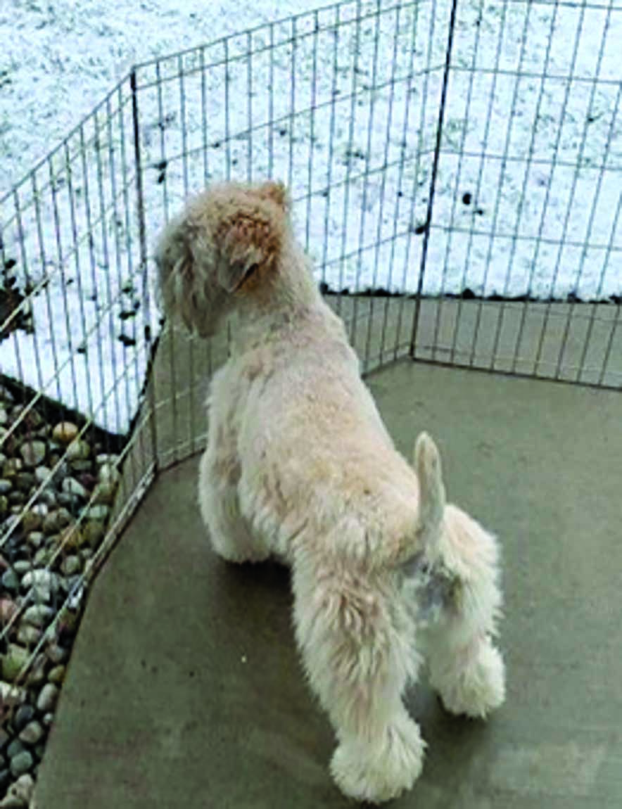
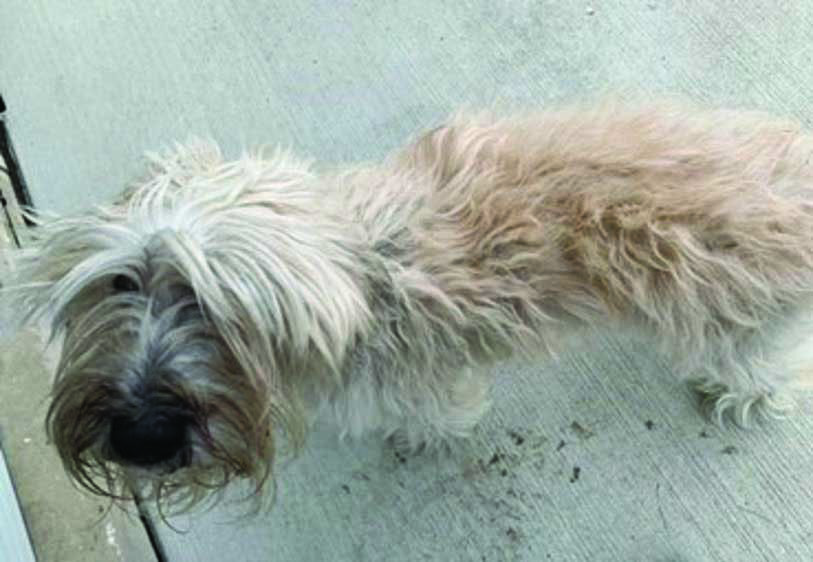
Physical examination findings are variable; in some cases, the exam is unremarkable, and in others severe deviations are present. Frequently observed abnormalities include decreased body condition and/or muscle due to malnutrition (Figure 1a & b), abdominal distension (and palpable “fluid-wave”), detection of peripheral edema, and/or decreased lung sounds secondary to pleural effusion. Chemosis secondary to hypoalbuminemia is seen rarely (Figure 2). Rectal examination may reveal thickened or roughened rectal mucosa and/or diarrheic stool.
|
|
| a |
 |
| b |
Figure 1. Images depicting the body condition of a 4-year-old, female neuter Soft-Coated Wheaten Terrier, before onset of clinical signs of PLE (a) and following diagnosis of PLE due to marked intestinal lymphangiectasia and moderate lymphoplasmacytic enteritis (b).
© Sara A. Jablonski
Hypoalbuminemia is the hallmark biochemical abnormality in cases of PLE. Additional common findings on complete blood count and serum biochemistry include lymphopenia, various types and degrees of leukocytosis, hypocholesterolemia, decreased serum creatinine, increased liver enzyme activities (typically mild increases), decreased total serum calcium and magnesium, and hypoglobulinemia. Although the latter is frequently observed, some dogs with PLE will have normal or even increased serum globulin concentrations.
Observation of serum total hypocalcemia is expected secondary to hypoalbuminemia, however ionized hypocalcemia also occurs, often associated with decreases in serum 25-hydroxyvitamin D. Concurrent ionized hypomagnesemia and secondary disorders of the parathyroid gland may also be present 6,7. Consideration should therefore be given to measurement of these variables. Hypocobalaminemia is also frequently observed in dogs with PLE, as are decreases in serum folate concentrations and increases in canine pancreatic lipase immunoreactivity (cPLI). Finally, viscoelastic testing has demonstrated a hypercoagulable state in dogs with PLE 8, but this finding has not been directly correlated to the development of thromboembolism.

Figure 2. Chemosis (a rare clinical consequence of hypoalbuminemia) in a 5-year-old male neuter Border Collie with PLE due to moderate lymphoplasmacytic and neutrophilic enteritis and mild intestinal lymphangiectasia.
© Sara A. Jablonski
The initial diagnostic work-up in cases of suspected PLE should involve careful consideration and exclusion of non-GI causes of hypoalbuminemia (Table 2). When necessary, measurement of fecal alpha 1-proteinase inhibitor can confirm that loss of protein through the gastrointestinal tract is occurring. Alpha 1-proteinase inhibitor is similar in size to albumin, and as it is not normally actively absorbed or secreted in the intestine, and is resistant to hydrolysis, it is an ideal marker for intestinal protein loss 9. This test is probably most useful in patients that have concurrent renal protein loss or liver dysfunction which complicates the diagnosis of PLE. Following this step, a variety of tests are recommended prior to intestinal biopsy. This includes screening for hypoadrenocorticism; a serum basal cortisol > 2 µg/dL rules it out, but if below this value ACTH stimulation testing should be performed to exclude hypoadrenocorticism. Other tests include fecal screening for helminths and Giardia duodenalis, diagnostic imaging, and specific testing for infectious diseases (e.g., urine antigen test and rectal scraping with cytology for histoplasmosis) depending on exposure and clinical suspicion.
Table 2. Non-GI causes of hypoalbuminemia and exclusionary tests.
| Disorder | Test(s) for exclusion |
|---|---|
| Liver insufficiency/dysfunction | Bile acids testing |
| Protein-losing nephropathy | Urinalysis +/- urine protein:creatinine ratio |
| Pancreatic insufficiency | Fasting serum trypsin-like immunoreactivity |
| Hemorrhage | Physical exam including rectal exam, evaluation for cavity effusions |
| Dilution or redistribution of albumin |
Evaluation for renal and cardiac disease
Evaluation for evidence of vasculitis or effusions
|
Thoracic radiographs may be useful to screen for evidence of pleural effusion, metastatic disease, or evidence of fungal disease. If chronic obstruction of the small intestine is a consideration, abdominal radiographs should be performed. Abdominal ultrasound may be helpful in excluding focal or extraluminal lesions that may change the diagnostic approach, and/or for fine-needle aspiration of masses or abnormal lymph nodes, which may allow a non-invasive diagnosis. Peritoneal effusion may be observed, and if present, fluid should be collected for analysis; a pure transudate is expected in cases of PLE. Hyperechoic mucosal striations (Figure 3) observed on abdominal ultrasound are supportive of, but not specific for, a diagnosis of intestinal lymphangiectasia 10. The diagnostic work-up should also include screening for abnormalities listed above – most importantly, ionized hypocalcemia and hypocobalaminemia.

Figure 3. Transverse ultrasound scan of small intestinal hyperechoic mucosal striations in a 7-year-old female neuter Goldendoodle with PLE.
© Sara A. Jablonski
Biopsy with histopathology is often required for definitive diagnosis of PLE, and remains an important step for several reasons. Primarily, biopsy can exclude infectious or neoplastic causes of PLE, but it will also help determine whether the dog is affected by CIE, IL or both (and if both, which process appears to predominate). Importantly, 76% of dogs with CIE and hypoalbuminemia will also have some degree of IL/lacteal dilation 11, so these processes are commonly present concurrently. Surgical exploration for biopsies provides the advantages of identifying focal areas of disease to biopsy and the ability to biopsy all segments of intestine, as well as other tissues as indicated (e.g., liver, lymph node). However, collection of intestinal tissue via flexible endoscopy offers many advantages and is generally the preferred option, as it is much less invasive and post-biopsy recovery is hastened when compared to laparotomy. Additionally, endoscopy enables direct visualization of the mucosa and allows for targeted collection of abnormal tissue. Specifically, “white spots” on the mucosa (Figure 4) have been associated with intestinal lymphangiectasia 12. Pathology can differ between intestinal sections, so it is highly recommended that both esophagogastroduodenoscopy (“upper”) and ileocolonoscopy (“lower”) endoscopy are performed 13. Importantly, endoscopy has limitations; the quality of endoscopic biopsies can impact the ability to make an accurate diagnosis, the jejunum typically cannot be sampled, and lesions deeper in the intestinal wall can be missed. Additionally, while guidelines for the interpretation of inflammatory and morphologic change in the GI mucosa of the dog and cat (WSAVA scoring system/template) are available 14, controversy and inter-observer variation exist in interpretation of intestinal biopsy samples. Additionally, histopathologic findings have not been consistently and accurately correlated to clinical signs and response to treatment. Some of the responsibility must be borne by the interpreting clinician to fully review the histopathology report, and to use their clinical judgment, particularly if samples are inadequate.

Figure 4. Presence of small intestinal pinpoint to coalescing “white spots” consistent with dilated lacteals in a 5-year-old female neuter Soft-Coated Wheaten Terrier with histologically diagnosed marked intestinal lymphangiectasia and the clinical syndrome of PLE.
© Sara A. Jablonski
Common histopathologic findings in dogs with PLE include intestinal lymphangiectasia (Figure 5), mucosal edema, various types and degrees of inflammatory infiltrates, and dilated, cystic crypts (Figure 6). Intestinal lymphangiectasia has been detected in the villus, proprial mucosa and submucosa in endoscopic intestinal biopsies 15, therefore it is important that pathologists evaluate for lymphangiectasia in each of these areas. Crypt lesions seem to be especially common in the Yorkshire Terrier 2. If concern for bacterial adherence/invasion arises from biopsy assessment, fluorescent in-situ hybridization (FISH) can be considered to evaluate for bacteria within formalin-fixed tissue. In some cases, immunohistochemistry and PCR for antigen receptor rearrangements (PARR) may be necessary to help distinguish intestinal lymphoma from inflammatory infiltrates.

Figure 5. Photomicrograph (x10 magnification) of marked intestinal lymphangiectasia and moderate lymphoplasmacytic, neutrophilic and eosinophilic duodenitis in a 5-year-old female neuter Soft-Coated Wheaten Terrier with PLE.
© Victoria Watson, DVM, PhD, Dip. ACVP

Figure 6. Photomicrograph (x40 magnification) of a markedly dilated crypt with degenerate inflammatory cells intermixed with eosinophilic necrotic debris and mucous in a 6-year-old, MC, small mixed breed dog with PLE.
© Victoria Watson, DVM, PhD, Dip. ACVP
The treatment of neoplastic, infectious, mechanical, and miscellaneous causes of PLE are beyond the scope of this review, and this section will focus on the treatment of PLE caused by CIE and IL. The severity of disease may influence the therapeutic approach to patients – so for dogs with suspected or confirmed PLE that are otherwise relatively stable, dietary treatment alone may be a reasonable approach. This approach has been shown to be successful in Yorkshire Terriers 16 and various other breeds 17. Additionally, it is critical to note that the treatment approach will often differ in individual cases of canine PLE, as it is a heterogenous disease process. In other words, there is no “cookbook” approach to treatment of PLE, and an individualized approach to therapy based on all available information is encouraged.
Although treatment should be directed towards the suspected or confirmed disease process, because PLE is a life-threatening disorder with a high rate of mortality, the safest approach may be to assume all processes (i.e., lymph fluid loss, increased intestinal permeability, mucosal injury) are occurring in a PLE patient, and treat accordingly. This is particularly true for patients with severe disease or those that are not responding to therapy.
Treatment of the underlying disease causing PLE starts with dietary modification, and many gastroenterologists consider this component of treatment to be the cornerstone of PLE management. One study suggests that dogs with PLE are more likely to respond to dietary therapy without the need for glucocorticoids if their canine chronic enteropathy clinical activity index (CCECAI) score is < 8 17. Dogs with PLE are in a catabolic state and may have marked negative energy and protein balance, so adequate nutrition is essential. Furthermore, treating PLE caused by CIE or IL relies on alterations to the diet. Anecdotally, the ideal diet is highly digestible, contains an adequate amount of protein, and is fat-restricted, but the dog’s previous dietary history should also be taken into consideration when choosing the best approach. A low-fat diet is typically recommended for dogs with IL, while a novel protein or hydrolyzed diet is suggested for dogs with CIE. Note there is no definitive consensus on what constitutes a “low-fat” diet in veterinary medicine; commercially available “low-fat” diets contain between 17-26 g fat/Mcal ME (1.7-2.6 g/100 kcal), while diets considered “ultra-low-fat” are typically classified as < 15 g fat/Mcal ME (1.5 g/100 kcal). Dogs that have IL as the cause of their PLE will often improve considerably with low fat diet alone, but some may require fat restriction beyond what a commercial diet can provide. Additionally, many of the commercially available diets lowest in fat are poultry based, which may make them unsuitable for dogs with IL that have concurrent CIE. At least one commercially available canned low-fat diet is currently pork-based, which may be novel for some dogs. Dogs that require fat restriction beyond what a commercial diet provides, and dogs that have both significant CIE and IL, may require a veterinary-nutritionist formulated, home-cooked diet that can address both concerns. In dogs with PLE and CIE and limited to no IL, a hydrolysate or novel protein diet can be considered, but it is still suggested to consider ones with comparatively lower fat content, because IL can be missed and serum albumin concentrations have been consistently correlated with lacteal lesions in dogs with inflammatory PLE 11,18. Other nutritional considerations include the food form (dry or wet), frequency of feeding (it is often beneficial to feed PLE dogs multiple, small meals a day), volume fed, and fiber content. Some dogs may benefit from supplementary fiber. In all cases of PLE, whether or not a commercially available therapeutic diet or a home-cooked one is desired or anticipated, consultation with a veterinary nutritionist is helpful and recommended.
Finally, it is important to recognize that lack of response to one dietary approach does not mean that the dog will not be diet-responsive, or that the condition will not benefit from optimization of the dietary therapy. In one study 8/10 dogs with steroid-refractory inflammatory PLE responded to a dietary change 19. In the author’s experience many dogs with PLE that have failed commercial diets and treatment with glucocorticoids and other immunosuppressive drugs have been rescued by feeding a novel, significantly fat-restricted (< 15% by ME), veterinary-nutritionist formulated, home-prepared diet. In some cases, dogs with PLE may not require a novel protein diet, but simply need fat restriction beyond what a commercial diet can offer, and so a home-prepared diet becomes necessary. A summary of specific diets for PLE conditions is given in Box 1.
Box 1. A summary of selected specific diets for PLE conditions.
| Intestinal lymphangiectasia: A veterinary therapeutic low-fat diet, or a home-cooked low-fat or ultra-low-fat diet formulated by a board-certified veterinary nutritionist. |
| Chronic inflammatory enteropathies: A veterinary therapeutic hydrolyzed or hypoallergenic diet, with priority given to those that are comparatively lower in fat, or a home-cooked diet formulated by board-certified veterinary nutritionist. |
| Combined lymphangiectasia and chronic inflammatory enteropathy: Hydrolyzed or hypoallergenic diets comparatively lower in fat could be considered, as can veterinary therapeutic low-fat diets. In some cases, management of one condition will allow for resolution of the other, but a home-cooked diet formulated by a veterinary nutritionist may need to be considered in cases where both disorders require dietary management. |
Although the pathogenesis of chronic inflammatory enteropathy is not completely understood, it is suspected that the GI tract has had a sustained immune reaction to endogenous or exogenous antigens (which may be food, bacterial and/or environmental). In addition, lymphangiectasia is associated with lymphangitis, and lymph leakage is known to induce secondary enteritis. Therefore, the initial approach to treating PLE typically involves the use of prednisone or prednisolone in either case. The exception may be in stable patients that are treated with diet alone initially and have shown a sustained clinical and biochemical response.
Importantly, side effects of steroid therapy in dogs with PLE can be significant, and glucocorticoids can worsen the catabolic and hypercoagulable states in some cases 20. Immunosuppressive doses of glucocorticoids may also be risky if a dog with PLE has a compromised enteric barrier, therefore in the author’s opinion it is important to carefully consider the dose of glucocorticoid being prescribed and use the most conservative dose that could be successful. Budesonide has a high first-pass effect and a high affinity for intestinal steroid receptors, and may be considered as an alternative glucocorticoid.
Some cases of PLE are started on an immunosuppressive drug at the time of diagnosis, or if response to appropriate doses of glucocorticoids is inadequate or side effects severe. It is important to note that there is no evidence of an immune process in cases of primary IL, thus immunosuppressive therapy is not warranted in these dogs. Furthermore, a recent study comparing time to normalization of albumin and long-term outcome between dogs with inflammatory PLE treated with steroids alone versus steroids plus a second-line immunosuppressive agent found no differences between the groups 21. Thus, the author recommends utilizing immunosuppressive agents (e.g., cyclosporine at 5 mg/kg PO q12-24H, or chlorambucil at 4-6 mg/m2 PO q24H for 7-14 days, then dose reduction) in patients that have CIE and are steroid-refractory, or in those that are initially steroid-responsive but relapse when steroids are weaned. A summary of the above is given in Box 2.
If both CIE and IL appear to be contributing to a patient’s PLE, it can be difficult to determine the best treatment approach, as one process may be driving the other. If IL is a significant component of the disease process it may be best to approach treatment for IL first and only escalate therapies if the patient fails to respond to therapies directed for IL.
Box 2. Drug recommendations for treating canine PLE.
|
Intestinal lymphangiectasia
|
|
Chronic inflammatory enteropathies
|
Dogs with PLE may develop an altered enteric microbiota (intestinal dysbiosis), thus probiotics may be helpful in some cases; at least one commercial probiotic strain has been shown to have a beneficial effect 22. As cobalamin is important for GI health and function, any deficiency should be treated; this has traditionally been administered subcutaneously, but recent work has demonstrated that oral administration can be effective in dogs with intestinal disease 23. Supplementation of folic acid should be considered in dogs with folate deficiency (200 µg/kg PO q24H – if < 20 kg and 400 µg/kg PO q24H – if > 20 kg), and human products are acceptable.
Treatment is recommended for dogs with significant ionized hypocalcemia. If clinical signs are observed (muscle twitching or tremors, facial rubbing), parenteral administration of 10% calcium gluconate may be necessary (0.5-1 mL/kg slowly IV over 10-30 minutes while monitoring heart rate, and optimally ECG). Oral calcium carbonate (25-50 mg/kg q24H, or elemental calcium at a divided dose q12H) can also be beneficial. It is important to remember that hypomagnesemia can impair calcium absorption, so if necessary oral magnesium hydroxide (1-2 mEq/kg q24H or as a divided dose q12H) can be given. Many dogs with ionized hypocalcemia have low 25-hydroxyvitamin D levels and may benefit from treatment with calcitriol (20-30 ng/kg PO q24H for the initial 3-4 days, followed by a maintenance dose of 5-15 ng/kg q24H, best given apart from steroids). It is unknown at this time whether dogs with PLE and hypovitaminosis D and normocalcemia would benefit from administration of vitamin D products. Dogs with PLE are classified as being at “high risk” for thrombosis (based on the 2022 CURATIVE guidelines) and thromboprophylaxis is recommended 24. Many dogs are administered clopidogrel at 2-3 mg/kg PO q24H, however the use of factor Xa inhibitors (e.g., apixaban, rivaroxaban) for thromboprophylaxis could also be considered.
Draining of abdominal or thoracic effusions is only recommended if there is discomfort or respiratory difficulty, use of diuretics is discouraged, as they are often ineffective and can promote dehydration. Any crystalloid fluid therapy should be judicious, due to the hypoproteinemia. The volume of plasma needed to increase a patient’s albumin is substantial and typically not practical. Colloids, such as hydroxyethyl starches, are the most useful option for improving edema. Concentrated human albumin (25%) is not recommended in dogs 25. A canine albumin product is available in some countries and is anecdotally effective in dogs with PLE. Finally, dogs with PLE can often benefit from other supportive care, such as medications to reduce vomiting and nausea (e.g., maropitant 2 mg/kg PO q24H).
If treating a dog without the benefit of an intestinal biopsy, a clinician should discuss with the client the risks involved (misdiagnosis, potential harms if the patient has an infectious enteropathy) and should also consider the breed involved and whether there are any known predispositions. In the absence of a biopsy or known breed predispositions, it may be best to assume that both IL and CIE are present, and treat accordingly.
Sara A. Jablonski
Some dogs with PLE have no to minimal clinical or biochemical response to anti-inflammatory or immunosuppressive doses of steroids and second-line immunosuppressive agents. In these cases, the author recommends tapering the medications and focusing on dietary adjustment (ideally with the consultation of a board-certified veterinary nutritionist), treating deficiencies, and preventing complications. Anecdotally, some dogs with refractory PLE caused by intestinal lymphangiectasia have shown improvement in response to octreotide (5-10 µg/kg SC q8-12H), but limited information about the efficacy and possible side effects of this treatment is currently available.
In a review of 445 cases of canine PLE, disease-associated death occurred in 54.2% of dogs 1. However, increasing understanding of the heterogenous nature of this condition and the need for individualized therapy may lead to better outcomes. Despite the guarded prognosis, some dogs with PLE will have an excellent response to treatment, but even in patients that respond initially, relapse is always possible. Affected dogs should be frequently monitored, and treatment can be life-long.
Protein-losing enteropathy (PLE) is a heterogenous syndrome in dogs most commonly caused by chronic inflammatory enteropathy, intestinal lymphangiectasia, or a combination of the two disorders. Diagnosis requires the exclusion of other causes of hypoalbuminemia, followed by a systematic work-up to identify the specific cause. Treatment should be individualized depending on the specific cause of a dog’s PLE, rather than a standardized approach, and dietary management is the cornerstone of therapy in many cases of canine PLE.
Craven MD, Washabau RJ. Comparative pathophysiology and management of protein-losing enteropathy. J. Vet. Intern. Med. 2019;33(2):383-402.
Simmerson SM, Armstrong PJ, Wünschmann A, et al. Clinical features, intestinal histopathology, and outcome in protein-losing enteropathy in Yorkshire Terrier dogs. J. Vet. Intern. Med. 2014;28(2):331-337.
Littman MP, Dambach DM, Vaden SL, et al. Familial protein-losing enteropathy and protein-losing nephropathy in Soft Coated Wheaten Terriers: 222 cases (1983-1997). J. Vet. Intern. Med. 2000;14(1):68-80.
Dossin O, Lavoué R. Protein-losing enteropathies in dogs. Vet. Clin. North Am. Small Anim. Pract. 2011;41(2):399-418.
Allenspach K, Iennarella-Servantez C. Canine protein losing enteropathies and systemic complications. Vet. Clin. North Am. Small Anim. Pract. 2021;51(1):111-122.
Allenspach K, Rizzo J, Jergens AE, et al. Hypovitaminosis D is associated with negative outcome in dogs with protein losing enteropathy: a retrospective study of 43 cases. BMC Vet. Res. 2017;8;13(1):96.
Jablonski Wennogle SA, Priestnall SL, Suárez-Bonnet A, et al. Comparison of clinical, clinicopathologic, and histologic variables in dogs with chronic inflammatory enteropathy and low or normal serum 25-hydroxycholecalciferol concentrations. J. Vet. Intern. Med. 2019;33(5):1995-2004.
Goodwin LV, Goggs R, Chan DL, et al. Hypercoagulability in dogs with protein-losing enteropathy. J. Vet. Intern. Med. 2011;25(2):273-277.
Heilmann RM, Parnell NK, Grützner N, et al. Serum and fecal canine α1-proteinase inhibitor concentrations reflect the severity of intestinal crypt abscesses and/or lacteal dilation in dogs. Vet. J. 2016;207:131-139.
Sutherland-Smith J, Penninck DG, Keating JH, et al. Ultrasonographic intestinal hyperechoic mucosal striations in dogs are associated with lacteal dilation. Vet. Radiol. Ultrasound. 2007;48(1):51-57.
Jablonski Wennogle SA, Priestnall SL, Webb CB. Histopathologic characteristics of intestinal biopsy samples from dogs with chronic inflammatory enteropathy with and without hypoalbuminemia. J. Vet. Intern. Med. 2017;31(2):371-376.
García-Sancho M, Sainz A, Villaescusa A, et al. White spots on the mucosal surface of the duodenum in dogs with lymphocytic plasmacytic enteritis. J. Vet. Sci. 2011;12(2):165-169.
Procoli F, Mõtsküla PF, Keyte SV, et al. Comparison of histopathologic findings in duodenal and ileal endoscopic biopsies in dogs with chronic small intestinal enteropathies. J. Vet. Intern. Med. 2013;27(2):268-274.
Washabau RJ, Day MJ, Willard MD, et al. Endoscopic, biopsy, and histopathologic guidelines for the evaluation of gastrointestinal inflammation in companion animals. J. Vet. Intern. Med. 2010;24(1):10-26.
Jablonski Wennogle SA, Priestnall SL, Suárez-Bonnet A, et al. Lymphatic endothelial cell immunohistochemical markers for evaluation of the intestinal lymphatic vasculature in dogs with chronic inflammatory enteropathy. J. Vet. Intern. Med. 2019;33(4):1669-1676.
Rudinsky AJ, Howard JP, Bishop MA, et al. Dietary management of presumptive protein-losing enteropathy in Yorkshire terriers. J. Small Anim. Pract. 2017;58(2):103-108.
Nagata N, Ohta H, Yokoyama N, et al. Clinical characteristics of dogs with food-responsive protein-losing enteropathy. J. Vet. Intern. Med. 2020;34(2):659-668.
Rossi G, Cerquetella M, Antonelli E, et al. The importance of histologic parameters of lacteal involvement in cases of canine lymphoplasmacytic enteritis. Gastroent. Hepatol. Bed. Bench. 2015;8(1):33-41.
Jablonski Wennogle SA, Stockman J, Webb CB. Prospective evaluation of a change in dietary therapy in dogs with steroid-resistant protein-losing enteropathy. J. Small Anim. Pract. 2021;62(9):756-764.
Jablonski Wennogle SA, Olver CS, Shropshire SB. Coagulation status, fibrinolysis, and platelet dynamics in dogs with chronic inflammatory enteropathy. J. Vet. Int. Med. 2021;35(2):892-901.
Salavati Schmitz S, Gow A, Bommer N, et al. Diagnostic features, treatment, and outcome of dogs with inflammatory protein-losing enteropathy. J. Vet. Intern. Med. 2019;33(5):2005-2013.
White R, Atherly T, Guard B, et al. Randomized, controlled trial evaluating the effect of multi-strain probiotic on the mucosal microbiota in canine idiopathic inflammatory bowel disease. Gut. Microbes. 2017;8(5):451-466.
Toresson L, Steiner JM, Suchodolski JS, et al. Oral cobalamin supplementation in dogs with chronic enteropathies and hypocobalaminemia. J. Vet. Intern. Med. 2016;30(1):101-107.
deLaforcade A, Bacek L, Blais MC, et al. 2022 Update of the Consensus on the Rational Use of Antithrombotics and Thrombolytics in Veterinary Critical Care (CURATIVE) Domain 1- Defining populations at risk. J. Vet. Emerg. Crit. Care (San Antonio) 2022;32(3):289-314.
Loyd KA, Cocayne CG, Cridland JM, et al. Retrospective evaluation of the administration of 25% human albumin to dogs with protein-losing enteropathy: 21 cases (2003-2013). J. Vet. Emerg. Crit. Care (San Antonio) 2016;26(4):587-592.
Sara A. Jablonski
Dr. Jablonski (previously Wennogle) received her DVM from Colorado State University (CSU) in 2011 Read more
Fecal microbiota transplantation is starting to be seen as a viable option to treat various acute and chronic gastrointestinal problems in dogs, as Linda Toresson explains.
Addison’s disease may not be the first diagnosis that comes to mind when a dog with gastrointestinal signs presents, but this possibility should not be dismissed, as Romy Heilmann describes.
Giardia infection is commonly identified in dogs, but deciding if it is a significant finding, and selecting the best treatment approach for a given situation, can often raise questions in practice; this article aims to provide some answers for the clinician.
There is now strong evidence that there are significant links between the gut and the kidneys, and that gastrointestinal health may be a key consideration when treating kidney disease, as discussed in this article